Pia Mater
The surfaces of the brain and spinal cord are lined with a firmly attached layer of pia mater. Unlike the arachnoid, the pia follows all contours of the brain and spinal cord and can not be removed with out damaging the underlying neural tissue. The pial layer creates a permeable border that allows interstitial fluid to pass from the brain to the surrounding cerebrospinal fluid in the subarachnoid space.
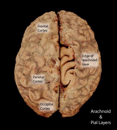 |
Figure: A superior view of a brain removed from the calvrium with arachnoid intact. In this view the arachnoid along the midline on the right cerebral hemisphere has been partial removal to expose the underlying pia mater that lines the surface of the brain. With the arachnoid gone the pia presents a smooth surface, |
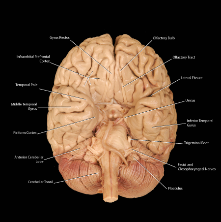 |
Figure: This is a ventral view of a brain with complete removal of the arachnoid layer to expose the pial surface. Since the pia is attached to the glial endfeet within the brain, the pia can not be removed with out damaging the surface of the brain. |
tr>
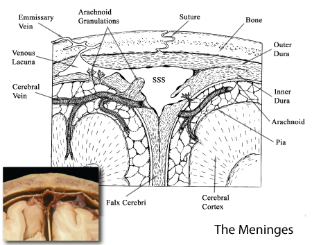 |
Figure: This is a summary diagram illustrating the pial surface as tightly adhered to the underlying cerebral cortical tissue. |