Spinal Meninges
| Spinal Dura | Arachnoid | Pia | Meningeal Spaces |
The spinal cord is surrounded by three layers of meningeal tissue: dura, arachnoid and pia. Although for the most part these three layers resemble the three layers of meninges present in the cranium, a significant difference however lies in the construction of the dural component. In the cranium the dura consists of two layers outer and inner dura closely associated with the periosteum of the cranial bones while in the spinal canal only the inner layer exists, termed the spinal dura and it is adherent to the posterior longitudinal ligament of the vertebral canal, not the periosteum of the vertebrae.
Spinal Dura Mater
The dural layer in the spinal canal is a sheet of dense connective tissue wrapped around the spinal cord continuously from the foramen magnum to the sacrum. Unlike the situation in the cranium where the dural mater is divided into two layers, the spinal dura is composed of only one layer and represents the spinal continuation of the inner layer of cranial dura. The spinal dura forms a tube into which the spinal cord is contained. Lateral extensions of the pia mater, termed denticulate ligaments, stabilize the spinal cord within the spinal dura. Distally the spinal dural tube closes at the level of the second sacral vertebrae (S2). From their it forms a thin filament of connective tissue that attaches to the coccygeal vertebrae.
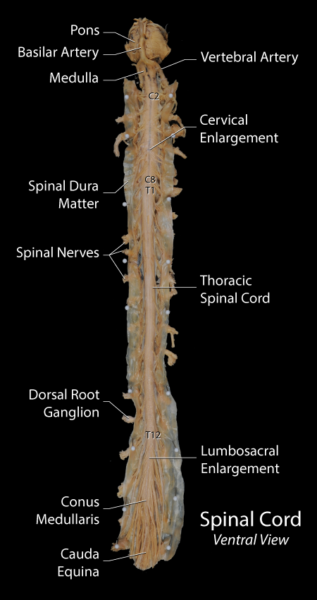 |
Figure: A ventral view of the spinal cord with the spinal dura opened to show its contents. The levels printed on the cord indicate root or segmental levels. The faint line in the center of the cord is the anterior spinal artery. |
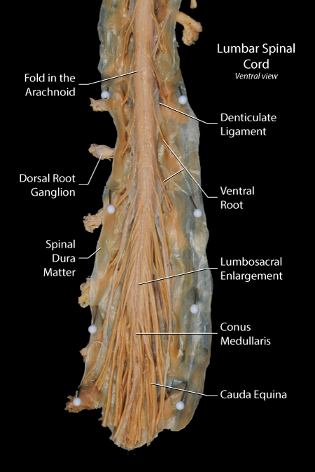 |
Figure: This is an ventral view of the lumbosacral region of the spinal cord following the opening of the dural and arachnoid to expose the spinal cord. |
| Spinal Dura | Arachnoid | Pia | Meningeal Spaces |
Arachnoid
The construction of the arachnoid is similar to that seen in the cranium. An outer membranous layer houses the cerebrospinal fluid and a complex network of tubercular extend from the membranous layer to the pia. Blood vessels and spinal roots pass through the matriculated portion. The blood vessels are surrounded by a sheath form from the trabeculae.
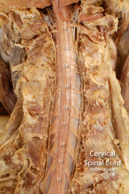 |
Figure: his is a posterior view of the post-laminectomy cervical spine. The laminae and spinous processes have been removed leaving the articular pillers on either side of the exposed spinal cord. The dural layer has been incised to expose the delicate arachnoid membrane covering the spinal cord and its roots. The dorsal roots are visible through the arachnoid. The board band coursing along the dorsal aspect of the cervical spinal cord (over which the cervical segments are numbered) represents the dorsal columns containing th. |
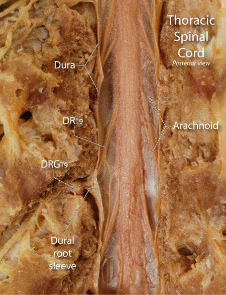 |
Figure: This is a posterior view of the post-laminectomy thoracic spine illustrating the meningeal relationship surrounding the formation of a thoracic spinal nerve. The dural layer has been incised to expose the delicate arachnoid layer covering the spinal cord and its roots. The T9 dorsal rootlets (DRT9) are seen joining together and entering a lateral recess to exit through an intervertebral foramen. The foramen has been unroofed to reveal the dorsal root ganglion of T9 (DRGT9). Note the small rootlet of T9 that communicates with a rootlet of T10 just before T9 enters the lateral recess. |
| Spinal Dura | Arachnoid | Pia | Meningeal Spaces |
Pia mater
The pial layer is tightly adherent to the underlying spinal cord. Close examination of the pia reveals that it is composed of several layers of delicate collagenous fibers with a squamous cell layer facing the cerebrospinal fluid. The arachnoid trabeculae join the pia. The space created between the membranous arachnoid and the pia, the subarachnoid space, houses the cerebrospinal fluid. Laterally, the pial layer extends off of the spinal cord as a thin, flatten sheet of connective tissue termed the denticulate ligament. The lateral border of this ligament forms a series of pointed extensions that anchor into the spinal dura. There is one attachment site in between each spinal root. In between these attachment the border of the ligament is scalloped creating a series of spaces between the ligament and the dura. it is through these spaces that the dorsal root passes to join with the ventral root and enter the lateral recess leading to the intervertebral foramen.
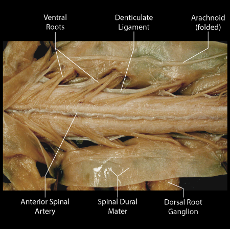 |
Figure: This is a ventral view of the cervical enlargement of the spinal cord with the dura incised to reveal the ventral roots and the anterior spinal artery. The pial layer is closely adherent to the surface of the spinal cord and can not be separated from the underlying spinal tissue. However, on the lateral margins of the spinal cord the pial layer extends outward as the denticulate ligament. |
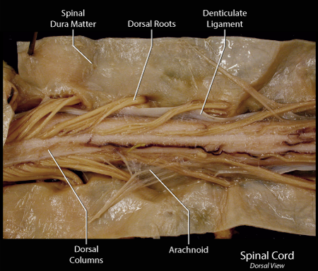 |
Figure: A dorsal view of the spinal cord with the dura incised to reveal the dorsal roots and the denticulate ligament. Superior is to the left and inferior to the right on this image. The dorsal columns represent an double column of white matter getting border as it progresses in a superior direction. Note the extension of the denticulate ligaments from the lateral margins of the spinal cord. |
| Spinal Dura | Arachnoid | Pia | Meningeal Spaces |
Meningeal Spaces
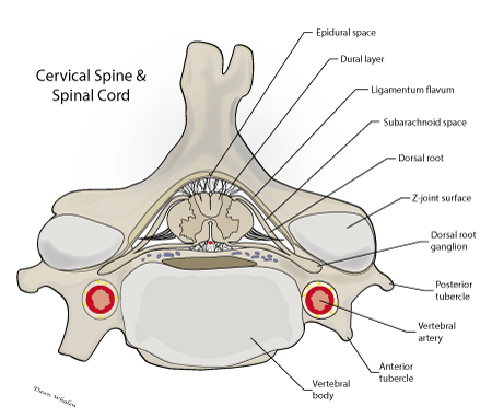
Two clinically significant spaces are created by the spinal meningeal layers: an epidural space located external to the spinal dura and a subarachnoid space located between the membranous arachnoid and the pia. The epidural space contains a small plexus of veins that extend the full length of the spinal canal, connecting at the superior end to the venous plexuses located in the cranium. At each vertebral level, the epidural plexus connects to the peripheral venous system through the intervertebral canal. The epidural space is also an important area for pain control, local anesthetics can be placed in the epidural space by cannula to anesthetize the spinal nerves as they enter the intervertebral canal. The subarachnoid space contains cerebrospinal fluid that bathes the nerve roots as they course through the spinal canal. Small spinal arachnoid granulations located just distal to the dorsal root ganglion allow for the absorption of CSF in to the epidural venous plexus.
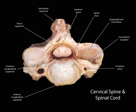 |
Figure: This is an axial plane section of the cervical spinal cord approximately at C3-4. It illustrates the structures surrounding the spinal cord. Since the individual was embalmed prone, the spinal cord is located in an abnormally dorsal position. A dural sheath is seen enveloping the cord and a slight epidural space separates the dura from the surrounding structures. The dorsal and ventral roots pass outward through the intervertebral canal or foramen located between the facet joint dorsally and the vertebral artery ventrally. The asterisk marks the dorsal root ganglion. |
|
Figure: This is a schematic diagram of an axial plane section of the cervical spine similar to the image presented above. Note the distinction between the epidural space, the dural layer and the subarachnoid space, |
| Spinal Dura | Arachnoid | Pia | Meningeal Spaces |