The Dura Mater
The outermost layer of meninges is the dura mater. It is composed of dense connective tissue. Cranial dura is actually composed of two tightly adhered layers: the outer dura or periosteal dura forms the periosteum on the inner aspect of the cranial bones. The inner dura or meningeal dura separates from the outer dura in several places to form the venous sinuses and the falx cerebri and tentorium cerebelli. The inner layer becomes the spinal dura when it passes through the foramen magnum.
The dural tissue is metabolically active and requires a decent source of blood, this is supplied by the meningeal arteries such as the middle and anterior meningeal arteries (shown below). These arteries course in between the two layers of dura. The dural is innervated by small meningeal nerves derived from the trigeminal nerve.
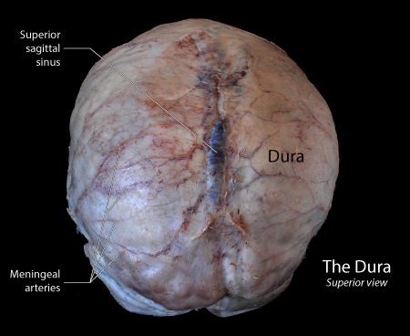 |
Figure: This is a view of the brain contained within a dural sac following the removal of the cranial bones. The visible surface is that of the outer dura or periosteum. The dark blue midline is created by the superior sagittal sinus. |
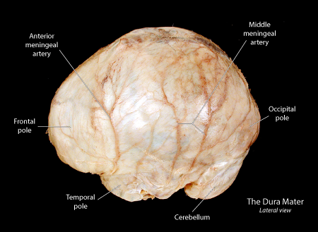 |
Figure: This is a lateral view of the brain covered with dura mate. The cranial dura mater is a thick layer of fibrous dense connective tissue that surrounds the brain, covering all three cranial fossae and continuous with the spinal meningeal dura over the foramen magnum, By lining the interior of the skull, the cranial dura represents a form of periosteum termed endosteum. |
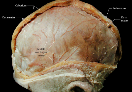 |
Figure: This is a lateral view of the head with left hemi-calvarium removed to expose the underlying dura. Sections of the middle meningeal artery can be seen on the surface of the outer dura. This artery is positioned between the outer dura and the bone of the calvarium. A small amount of periosteum is present on the outer surface of the calvarium (right side); the dura represents the periosteum (also termed endosteum) on the inside of the calvarium. |
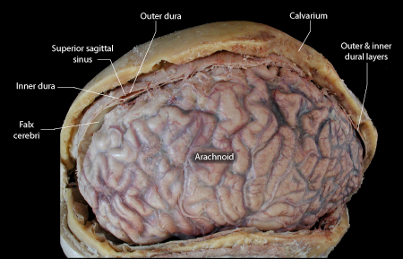 |
Figure: This image demonstrates the arachnoid following removal of the dura. The arachnoid is flattened onto the brain due to the loss of under-lying cerebrospinal fluids after death. The cut edge of the dura reveals the outer and inner layers separating to expose the superior sagittal sinus. |
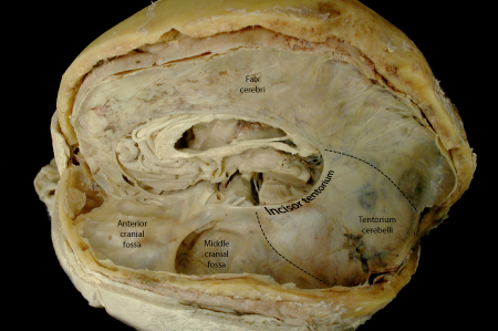 |
Figure: This image presents the cranial cavity after hemispherectomy. The falx cerebri is seen on the midline. The dashed line at the posterior aspect of the falx marks its border with the tentorium cerebelli. The second dashed line marks the attachment of the tentorium to the petrous ridge of the temporal bone. The incisor of the tentorium is the free border of the tentorium. |
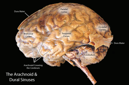 |
Figure: This is a lateral view of the brain with the dural layer mostly removed. The membranous arachnoid is exposed over most of the surface of the brain. A strip of dura mater has been left over the superior sagittal sinus and the transverse sinuses. The latter dura can be seen forming the transverse sinus and giving rise to the tentorium cerebelli, a plate of inner dura which separates the cerebellum from the occipitotemporal cortex. |
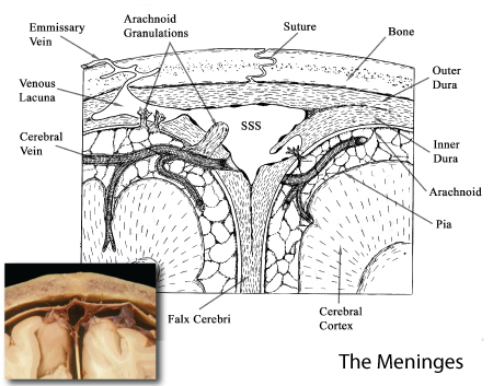 |
Figure: This is an illustration of a cross-section of the meninges taken through a sinus to illustrate the formation of dural sinuses. An outer and inner layer of dura is seen with the inner layer separating to form a dural sinus. A membranous arachnoid is seen under the inner dural layer and trabeculae extend inward to reach the pial layer on the surface of the brain. |