Spinal Meninges Dissection
1084BI
This dissection is a posterior approach to the spinal canal and spinal cord. It begins following the removal of the paraspinal muscles and a complete laminectomy to expose the spinal dura. In the cervical region the dura is opened to expose the arachnoid forming the cisterna magna after which the arachnoid is removed and the relationships around the C2 nerve root are examined. Similarly the dura is opened in the thoracic spine, exposing the arachnoid which is then removed and the relationships around the T9 nerve root are examined.
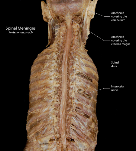 |
Figure: This is a posterior view of the cervical and thoracic vertebral column following the removal of the paraspinal muscles and a complete laminectomy to expose the spinal dura. Superiorly, the occipital bone has been removed and the dura has been opened to expose the underlying arachnoid. |
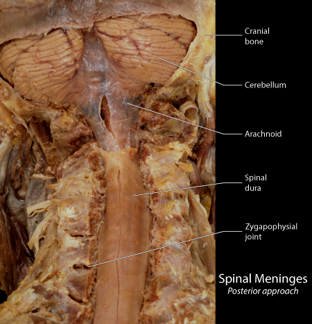 |
Figure:The dura has been opened to expose the arachnoid layer over the cerebellum and through the foramen magnum to the level of the C2 vertebra. A small slit was placed in the arachnoid at the level of the cisterna magna to expose the inside of the cistern. |
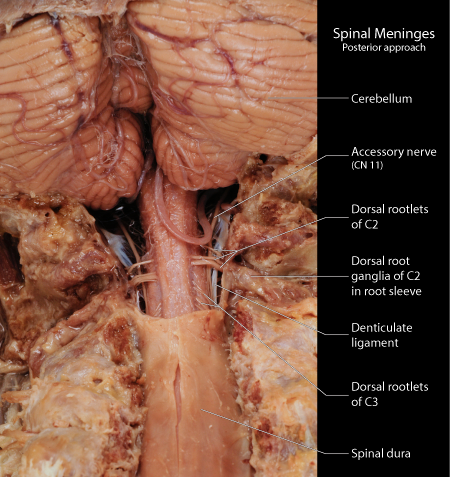 |
Figure: In this view the arachnoid has been removed to expose the |
 |
Figure: |
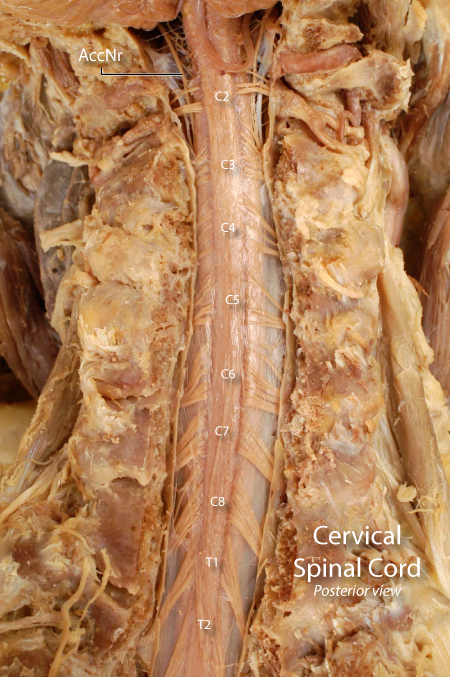 |
Figure: |
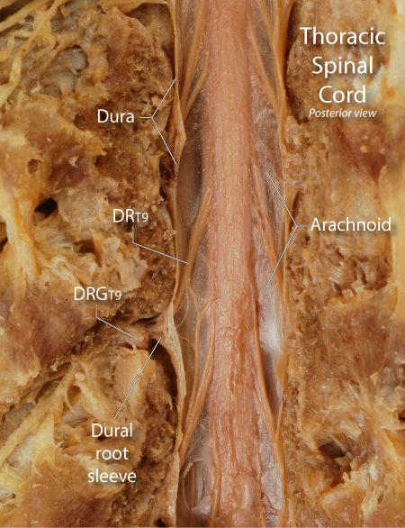 |
Figure: |
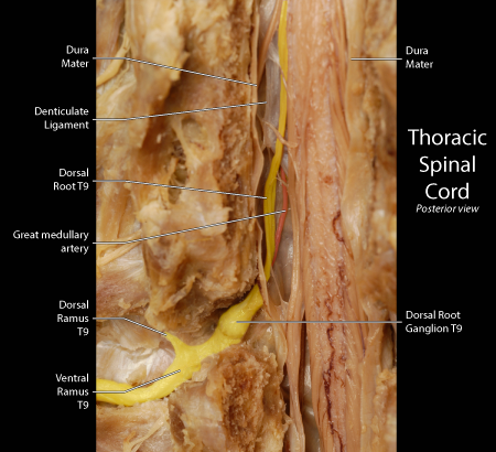 |
Figure: |