The Calvarial Floor
During development the brain becomes ensheathed in a membrane, the primitive meninges (meninx). The outermost layer of the primitive meninges differentiates into dura and has the potential to form bone. At this stage the brain is said to housed in the desmocranium. The portions of the membrane located ventral to the brain come under compression and rapidly convert to cartilage forming a chondocranium. The cartilage, through a process of endochondrial ossification, converts to bone forming the basicranium. The portions of the desmocranium surrounding the superior portion of the brain become stretched with the rapid growth of the cerebrum and begin forming bone directly through a process of intramembranous ossification.
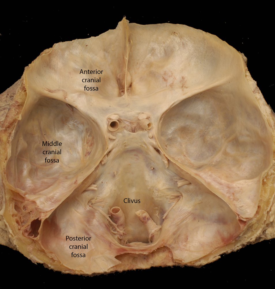
During the 2nd and 3rd months of development the bone is just beginning to form and is easily molded by the growing brain. At this time the ventral surface of the brain makes three notable impressions on the floor of the calvarial: the anterior, middle and posterior cranial fossae. Each fossa is associated with a specific portion of the brain.
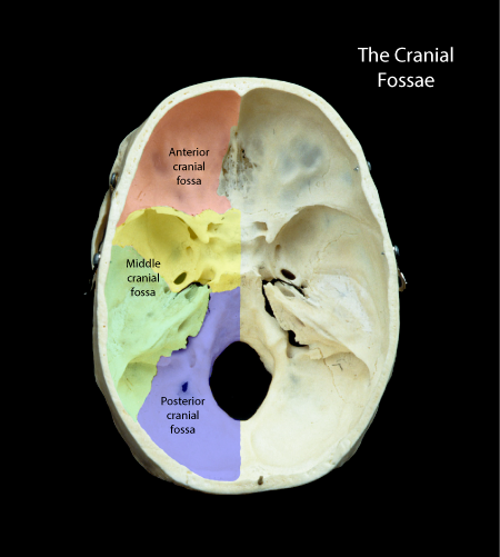 |
Figure: This is a view of the floor of the calvarium. The major bone making up the calvarial floor have been color coded. (Red, Frontal bone; Yellow, Sphenoid bone; Green, Temporal bone; Blue, Occipital bone. |
The anterior cranial fossa houses the ventral aspect of the frontal lobes of the brain. The middle cranial fossa contains much of the temporal lobe and the posterior cranial fossa surrounds the brainstem and cerebellum.
As the bone of the cranial base is forming, it condenses around the cranial nerves as they exit the brainstem and enter the visceral portion of the cranium. This condensation creates a number of foramina that transmit the cranial nerves. Shown below are two preparations where the occipital bone was sectioned, the entire brain removed but the roots of the cranial nerves have remained in their respective foramina.
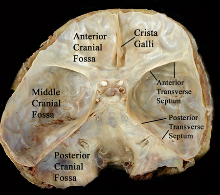 |
Figure: A view of the calvarial floor covered with dura mater. The occipital bone was sectioned in order to remove the brainstem and cerebellum. |
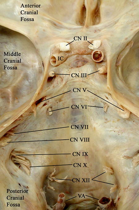 |
Figure: This is a magnified view of the above image illustrating the roots of the cranial nerves. Cranial Nerve II enters the orbital canal and Cranial Nerve III, IV, and VI enter the cavernous sinus. Cranial Nerve V has three components: V1 enters the cavernous sinus, V2 enters the foramen rotundum and V3 enters the foramen ovale. Cranial Nerves VII and VIII leave the cranial cavity through the internal acoustic meatus while Cranial Nerves IX, X and XI exit through the jugular foramen. Finally Cranial Nerve XII leaves through the hypoglossal canal. |
 |
Figure: The cranial floor exposed similar to the above images. |
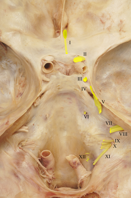 |
Figure: A magnified image of the above dissection with the roots of the cranial nerves indicated in color. |