The Dural Venous Sinuses
The cranial dura mater is composed of two layers, outer and inner. The outer layer repesents the periosteum of the cranial bones while the inner layer represents meningeal dura. The inner layer separates from the outer layer in several locations to form a dural venous sinus. At these locations the two layers of inner dural reunite to form a dural partition such as the falx cerebri and the tentorium cerebelli.
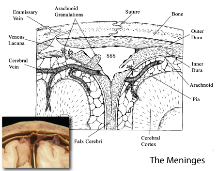 |
Figure: This is an illustration of a cross-section of the meninges taken through a sinus to illustrate the formation of dural sinuses. An outer and inner layer of dura is seen with the inner layer separating to form a dural sinus. A membranous arachnoid is seen under the inner dural layer and trabeculae extend inward to reach the pial layer on the surface of the brain. |
Each of the dural venous sinuses is ineffect a vein, the internal walls of the sinus are lined with a vascular endothelium. The dural venous sinuses collect blood from the cerebral veins. The venous blood is then brought to the jugular foramen where it is delivered to the beginning of the jugular vein. These hard-walled sinuses also serve as collecting vessels for cerebrospinal fluid (CSF). The underlying arachnoid forms small one-way valves, termed arachnoid granulations, that protrude into the dural sinus allowing for the movement of CSF from the subarachnoid spaces into the venous sinus.
 |
Figure: This is a superior view of the brain following the removal of all dura except that covering the dural sinuses. The dura covering the superior sagittal sinus is seen in this image. Not visible on this image are a series of small arachnoid granulations extending into the under surface of the superior sagittal sinus. |
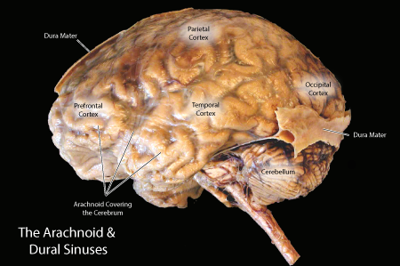 |
Figure: This is a lateral view of a brain with the dura removed except for the regions inwhich it forms a sinus. The dura covering the transverse sinus is seen in this view. The transverse sinus has been sectioned to demonstrate its construction from the two sheets of inner dura. |
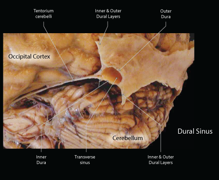 |
Figure: This is a magnified view of the image above demonstrating the three walls of the transverse sinus. The outer dura forms the posterior wall while the two layers of inner dura form the superior and inferior walls. Both layers of inner dura unite to form the tentorium cerebelli, which separates the occipitotemporal lobes from the cerebellum. |
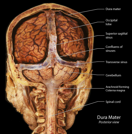 |
Figure: This is a posterior view of a cranium of an 80-year-old female following the removal of the posterior portion of the calvarium. The dura mater has also been removed except where it overlies the venous sinuses. |
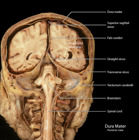 |
Figure: This is the same sopecimen as above following the removal of the posterior portion of the cerebral hemispheres. In this preparation, four dural venous sinuses have been sectioned in the coronal plane: superior sagittal sinus, straight sinus, and the left and right transverse sinuses. |
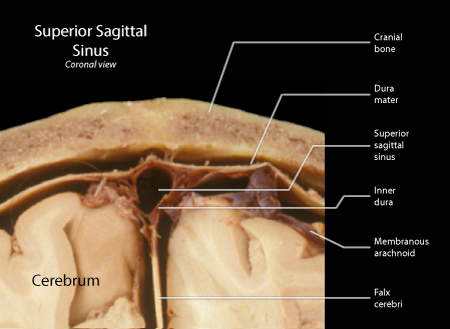 |
Figure: Magnified view of the superior sagittal sinus from the above preparation. |
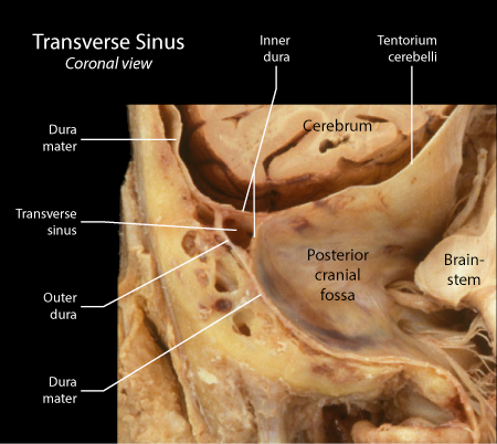 |
Figure: Magnified view of the left transversse sinus from the above preparation. Note the separation of the two layers of inner dura to make the superior and inferior walls of the transverse sinus and the union of these two layers to form the tentorium cerebelli. |
The dural venous sinuses represent the major drainage route for blood from the brain. Sveral of the sinuses are of critical importance, these are: the superior and inferior sagittal sinuses draining the cerebrum, the straight sinus draining the sagittal sinuses and the great vein of Galen, the transverse and sigmoid sinuses draining the confluens of sinuses (trigone) and the petrosal sinuses draining the temporal bone and posterior cranial fossa.
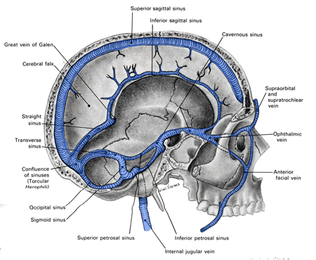 |
Figure: |