The Dural Partitions
| Falx Cerebri | Tentorium cerebelli | Anterior Dural Girdle | Diaphragma Sella |

This is a lateral view of the head with the brain removed to illustrate the dural partitions. (Figure taken from: Thorek., P., Anatomy in surgery, New York:Springer-Verlag, 1985.
The inner layer of dura expands internally to form a series of five partitions that separate portions of the brain. On the midline, the inner dural layers separate around the superior sagittal sinus and then unite to form the falx cerebri also termed the median dural septum, which lies between the two cerebral hemispheres. Laterally the inner dural layers separate to form the transverse sinus and then unite to form the tentorium cerebelli also termed the posterior dural girdle, which separates the cerebellum from the cerebrum. A small partition arises from the under surface of the tentorium and is termed the falx cerebelli, it partially separates the two cerebellar hemispheres. In addition to these three dural partitions a fourth partition arises from the posterior margin of the lesser wing of the sphenoid. This partition, termed the anterior dural girdle, separates the temporal lobe from the frontal lobe. Finally, the inner dura forms a fold the partially covers the sella turcica termed the diaphragma sella. This partition separates the pituitary from the base of the brain.
The Falx Cerebri
The falx cerebri is a cycle-shaped sheet of inner dura that separates the two cerebral hemispheres. Anteriorly it is attached to the crista galli of the ethmoid bone while posteriorly the inner dural layers separate around the straight sinus to help form the tentorium cerebelli. The superior surface of the falx cerebri splits to form the superior sagittal sinus and attaches to the outer dura. The inferior or free border of the falx contains the inferior sagittal sinus and forms sharp edge that course just above the corpus callosum.
 |
Figure:
|
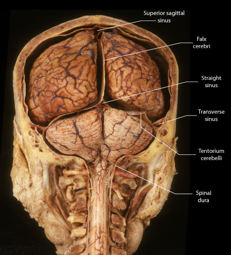 |
Figure: |
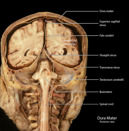 |
Figure: |
| Falx Cerebri | Tentorium cerebelli | Anterior Dural Girdle | Diaphragma Sella |
Tentorium Cerebelli
When the flax cerebri divides to form the straight sinus the two walls of the dural sinus continue laterally to contribute to the tentorium cerebelli. Laterally the tentorium splits to form the superior petrosal sinus and to attach to the petrous ridge. Posteriorly the tentorium splits to form the transverse sinus and attach to a groove on the inner aspect of the occipital bone. Anteriorly the tentorium has a free border that forms a sharp edge termed the incisor of the tentorium. The incisor is wrapped closely around the brainstem. The tentorium separates the cerebellum in the posterior cranial fossa from the cerebrum, thus this latter structure is said to have a supratentorial location.
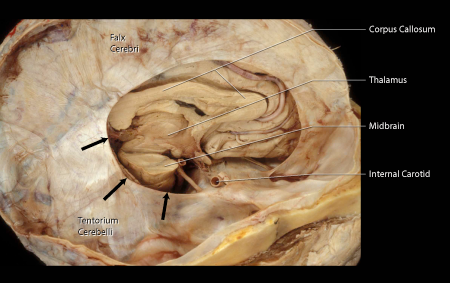 |
Figure: This is a photograph of a dissection illustrating the tentorium cerebelli and its cutting edge - the incisor tentorium. The right hemisphere of the cerebrum has been removed to reveal the continuity between the tentorium and the falx cerebri. The free border of the tentorium is sharp and referred to as the "cutting edge" or incisor tentorium, also termed the tentorial incisure (black arrows). |
|
Figure: This is a posterior view of the cranium sectioned in the coronal plane. Note that the separation of the cranial dura from the calvarium is an artifact of this preparation. The cranial dura can be seen forming the superior sagittal sinus and the transverse sinus. From these two sinuses, the falx cerebri and the tentorium cerebelli are formed. The falx cerebri and the tentorium cerebelli then unite on midline to form the straight sinus. |
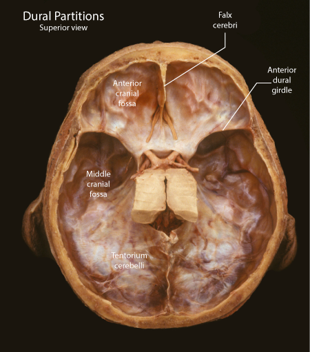 |
Figure: This is a superior view of the calvarium with the cerebrum removed to demonstrate the anterior and middle cranial fossae. The tentorium cerebelli covers the posterior cranial fossa. The tentorium rises toward the midline where it forms the falx cerebri, which has been sectioned in order to remove the cerebrum. The frontal lobes sit in the anterior cranial fossa, the temporal lobes lie in the middle cranial fossa and the occipital lobes are positioned on the tentorium. |
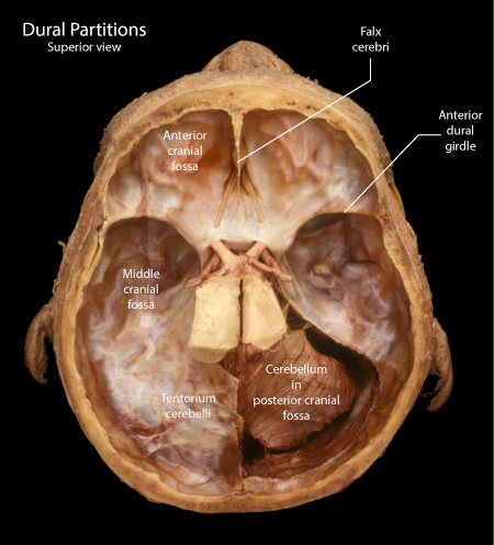 |
Figure: In this view of the same specimen as above the right tentorium has been removed to reveal the underlying posterior cranial fossa containing the cerebellum and brainstem. The cut surface of the tentorium attaches to the petrous ridge of the temporal bone. |
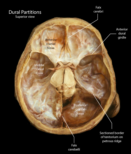 |
Figure: In the third view of the same specimen, the cerebellum has been removed to illustrate the extent of the posterior cranial fossa. The posterolateral surface of the brainstem is visible in the posterior cranial fossa. Note also cranial nerves seven and eight leaving the lateral aspect of the brainstem and entering the internal auditory meatus on the medial surface of the temporal bone |
| Falx Cerebri | Tentorium cerebelli | Anterior Dural Girdle | Diaphragma Sella |
Anterior Dural Girdle
The dura covering the inferior border of the lesser wing of the sphenoid bone forms a band that extends laterally onto the wall of the calvarium. The ridge so formed is termed the anterior dural girdle. It separates the temporal lobe from the frontal lobe. The posterior and lateral extension of the girdle helps to create a recess in to which the temporal pole is inserted. Rapid changes in the velocity of the head can result in this girdle severely injuring the temporal pole.
|
Figure: This is a superior view of the calvarium with the cerebrum removed to demonstrate the anterior and middle cranial fossae. The tentorium cerebelli covers the posterior cranial fossa. The anterior dural girdle is seen as a sharp ridge coursing along the posterior margin of the lesser wing of the sphenoid bone. |
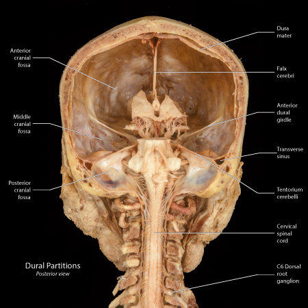 |
Figure: This is a posterior view of the calvarium following the removal of the cerebrum and cerebellum. The falx cerebri is seen on the midline, the anterior dural girdle is seen forming the roof of the middle cranial fossa and the tentorium has been sectioned along the edge of the petrous ridge. |
| Falx Cerebri | Tentorium cerebelli | Anterior Dural Girdle | Diaphragma Sella |
The Diaphragma Sella
The opening to the sella turcica is partially covered by a fold of dura termed the diaphargma sella. The stalk passes through the diaphragm to reach the pituitary. Cerebrospinal fluid can also pass through this opening in the diaphragma sella. Occlusion of this circulation can raise the pressure in the sella turcica and compress the pituitary. On imaging the pituitary is flattened against the walls of the sella and not visible, but may be still functioning. This condition is termed "Empty Sella Syndrome".
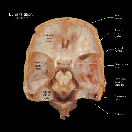 |
Figure: This is a superior view of the calvarium following the removal of the brain. In the center of the calvarium is the sella turcica covered by a fold in the dura termed the diaphragma sella. |
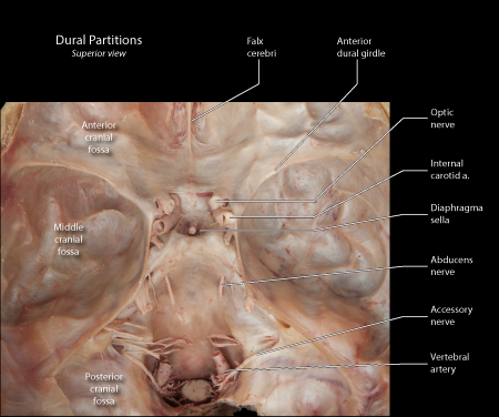 |
Figure: This is a superior view of the calvarium following the removal of the brain. Note the infundibulum (pituitary stalk) in the center of the diaphragma sell. |