Cerebellar Anatomy
The cerebellum or "little brain" lies in the posterior cranial fossia stradling the posterior aspect of the fourth ventricle of the brainstem. It is composed of two large hemispheres positioned laterally and a medially located vermis.
The head in the Figure One was sectioned in the sagittal plane approximately on the midline. In this view, only the midline vermis of the cerebellum can be seen. Note its relationship to the braintem and fourth ventricle.
The cerebellum is surrounded superiorly by the tentorim cerebelli and the superior cerebellar cistern (Figure Two). The inferior border consists of the inferior cerebellar cistern or cisterna magna and the foramen magnum. Posteriorly, the cerebellum is bounded by the tentorium and the occipital bone while the anterior border is composed of the fourth ventricle and brainstem. Three large fiber tracts, termed peduncles, attach the cerebellum to the brainstem. The superior cerebellar peduncle attaches to the midbrain, the middle cerebellar peduncle to the pons and the inferior cerebellar peduncle to the medulla.
The head in Figure Three was prepared by removing the occipital bone and portions of the parietal bones. It demonstrates the two hemispheres of the cerebellum in the posterior cranial fossa. The superior sagittal and transverese sinuses have been left in place. Note the thin velum of arachnoid drapped off the inferior aspect of the cerebellum and extending down through the foramen magnums to surround the spinal cord in the spinal canal. The space encased by the arachnoid is the cisterna magna or inferior cerebellar custern.
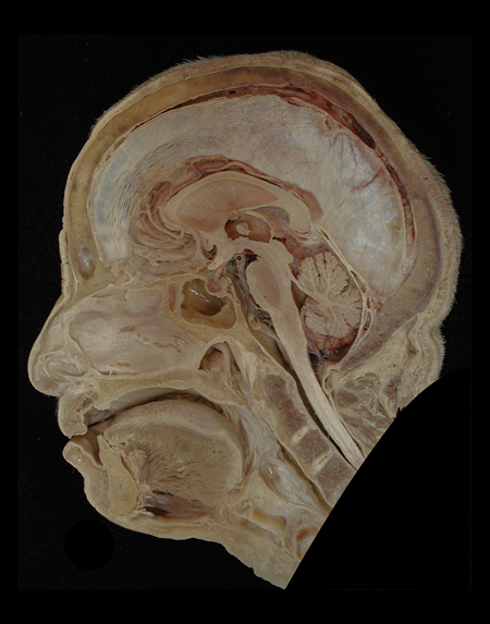 |
Figure: A midsagittal view of a male head illustrating the location of the cerebellum in the posteror cranial fossa. |