Overview of Cerebral Gyri
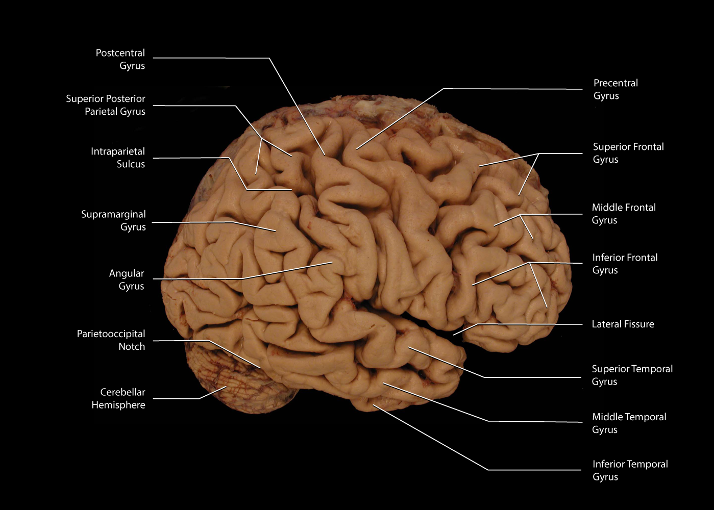
The cerebrum is composed of a massive collect of white matter tracts covered by a thin mantle of gray matter. The white matter is arranged in reasonably consistent folds creating a series of gyri and sulsi on the surface of the cerebral cortex. The gyral pattern of the cerebrum has been associated with specific aspects of cortical function.
When examining any one brain, the pattern of gyri may appear to be variant, however there is a generalized pattern into which all brains tend to fit:
Frontal lobe
Three major frontal gyri - superior, middle and inferior - that are oriented parallel to the longitudinal fissure separating the hemispheres
Parietal lobe
A postcentral gyrus oriented orthogonal to the longitudinal fissure and the lateral issue and superior and inferior parietal lobules separated by an interparietal sulcus oriented parallel to the longitudinal fissure
Temporal lobe
Three major temporal gyri - superior, middle and inferior - that are oriented parallel to the long axis of the temporal lobe
Occipital lobe
A calcarine sulcus separating two gyri on the medial surface of the brain
Insula lobe
A series of several gyri oriented orthogonal to the lateral fissure and positioned deep withing the lateral fissure.
This page will present a number of labeled views of the cerebrum from several different views. These are:
| Lateral views | Superior views | Inferior views | Frontal views | Posterior views | Ventral views | Medial views |
Image Library
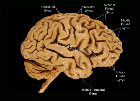 |
Figure: |
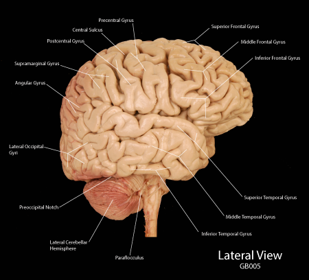 |
Figure: |
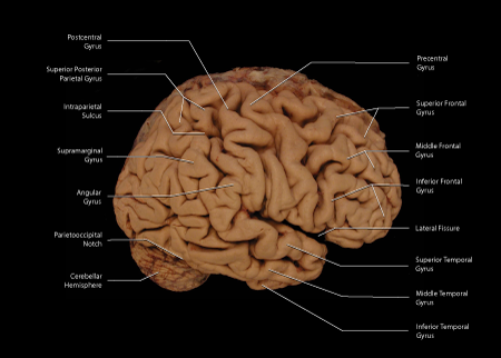 |
Figure: |
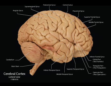 |
Figure: |
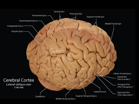 |
Figure: |
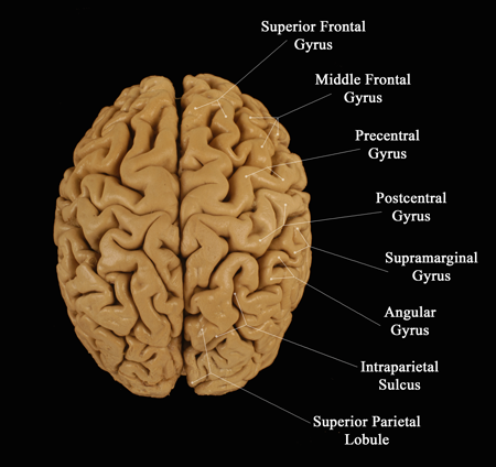 |
Figure: |
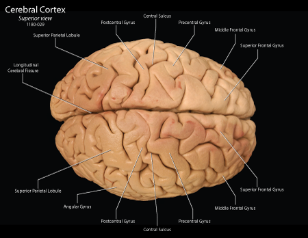 |
Figure: |
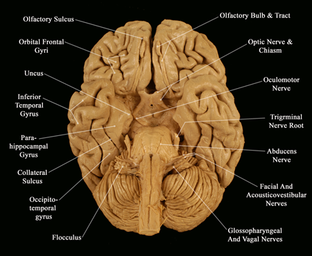 |
Figure: |
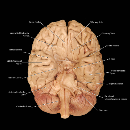 |
Figure: |
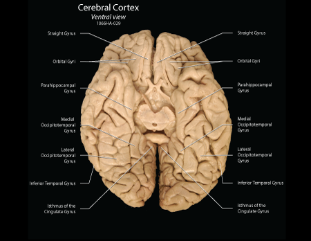 |
Figure: |
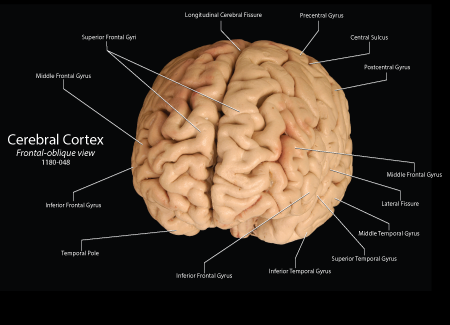 |
Figure: |
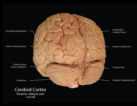 |
Figure: |
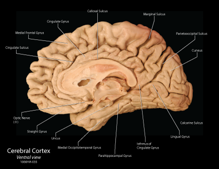 |
Figure: |
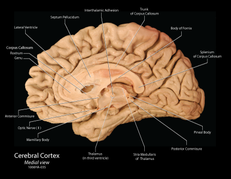 |
Figure: |
| Figure:
Enlarge image in new window |