The Arachnoid
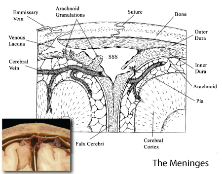
The arachnoid forms a delicated layer loosely attached to the undersurface of the dura. It is composed of two parts, a continuous membranous portion that is every where attached to the inner dural surface and a trabeculated portion that links the arachnoid to the underlying pial layer. The arachnoid covers the surface of the brain and extends through the foramen magnum to also surround the spinal cord eventually reaching the inferior tip of the dural sac at the S2 vertebral level.. Over most of the surface of the brain the arachnoid follows the cerebral contours, however in several regions this membranous structure leaves the surface of one region of the brain to pass onto another rregion. Where this happens a subarachnoid cistern, filled with cerebrospinal fluid (CSF), is created.
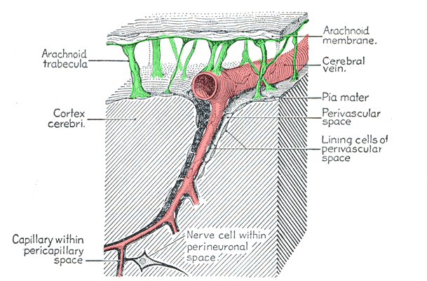
A diagram illustrating the presence of the verebral arteries in the subarachnoid space and the extension of this space along the margin of the penetrating vessels as the space of Virchow-Robbins. (Figure taken from: Ranson,S.W. and Clark,S.L., The Anatomy of the Nervous System, W.B. Saunders Co., Philadelphia, 1961.)
The arteries supplying the cerebrum course through the subarachnoid space wraped in a layer created by the trabecular arachnoid. The pial layer follows the penetrating vessels into the cerebral tissue creating an extension of the subarachnoid space along the wall of the vessel. This is termed the perivascular spcae or Virchow-Robbins space and its continuity with the subarachnoid space allows cerebrospinal fluid the surround the penetrating vessels.
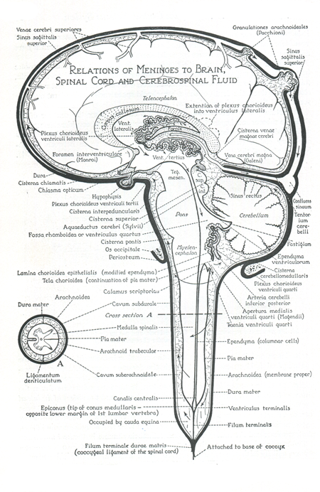 |
Figure: This is a schematic diagram illustrating the subarachnoid spaces surrounding the brain and spinal cord. Arachnoid granulations returning CSF to the venous system are seen in the superior sagittal sinus. Similar granulations (not shown here) are also present in the dural root sleeve of each spinal nerve. (Figure taken from: Ranson,S.W. and Clark,S.L., The Anatomy of the Nervous System, W.B. Saunders Co., Philadelphia, 1961.) |
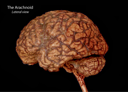 |
Figure: A lateral view of a human brain that was removed with the arachnoid layer intact. Note the presence of blood vessels covered with arachnoid on the surface of the cerebrum and cerebellum. |
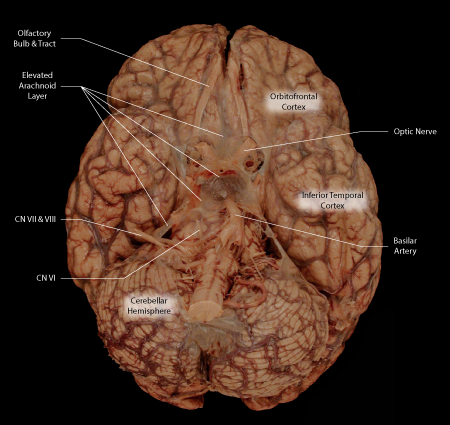 |
Figure: A ventral view of the same specimen as shown above. Along the ventral midline, anterior to the brainstem as well as inferior to the cerebellum, the membranous arachnoid does not follow the contours of the brain thus creating enlargements of the subarachnoid space termed cisterns. These arachnoid cisterns are filled with CSF as is the rest of the subarachnoid space. |
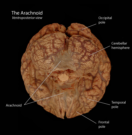 |
Figure: A ventroposterior view of the same specimen as above illustrating the membranous arachnoid overlying the cisterna magna or inferior cerebellar cistern that lies inferior to the cerebellum and the arachnoid overlying the pentangular cistern that lies anterior to the brainstem. |
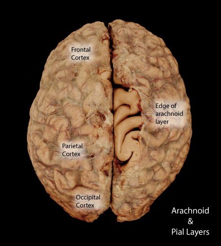 |
Figure: A superior view of the same specimen as above illustrating the partial removal of the arachnoid to expose the underlying pia mater that lines the surface of the brain. |
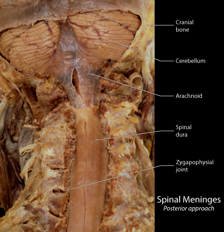 |
Figure: This is a posterior view of the craniocervical junction illustrating the continuity of the membranous arachnoid from the inferior aspect of the cerebellum into the spinal canal. A small slit in the membranous arachnoid reveals the underlying cisterna magna. |
