The Meninges: Microarchitecture
The central nervous system is surrounded by three separate layers of meninges: dura, arachnoid and pia. The dural layer is closely related to the periosteum of the surrounding bones while the remaining two layers, termed leptomeninges, are closely related to the brain and spinal cord.
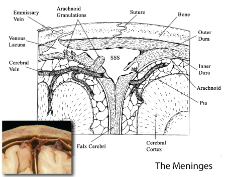 |
Figure: A schamtic diagram of the three meningeal layers as seen in the cranium. The drawing is derived from the meningeal relationships illustrated in the inset, a photograph of the superior sagittal sinus and associated meninges sectioned in the coronal or frontal plane. |
Dura mater
The dura is composed of dense connective tissue containing collagen bundles and fibrocytes. In the cranium, the dural layer is divided into two portions: the outer dura or endosteum of the cranial bone and the inner dura. The microarchitectural composition of both layers is similar. In the spinal canal, the spinal cord is surrounded by only one layer of dura, often termed spinal dura. Its microarchitectureal composition is similar to that of the inner dura of the cranium. The spinal dura evaginates to follow the spinal roots to a point just distal to the dorsal root ganglion. Here the dural layer transforms into the epineurium of the peripheral nerve. The lateral extention of the dura surrounding the spinal roots is often termed the root sheath.
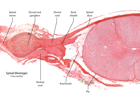 |
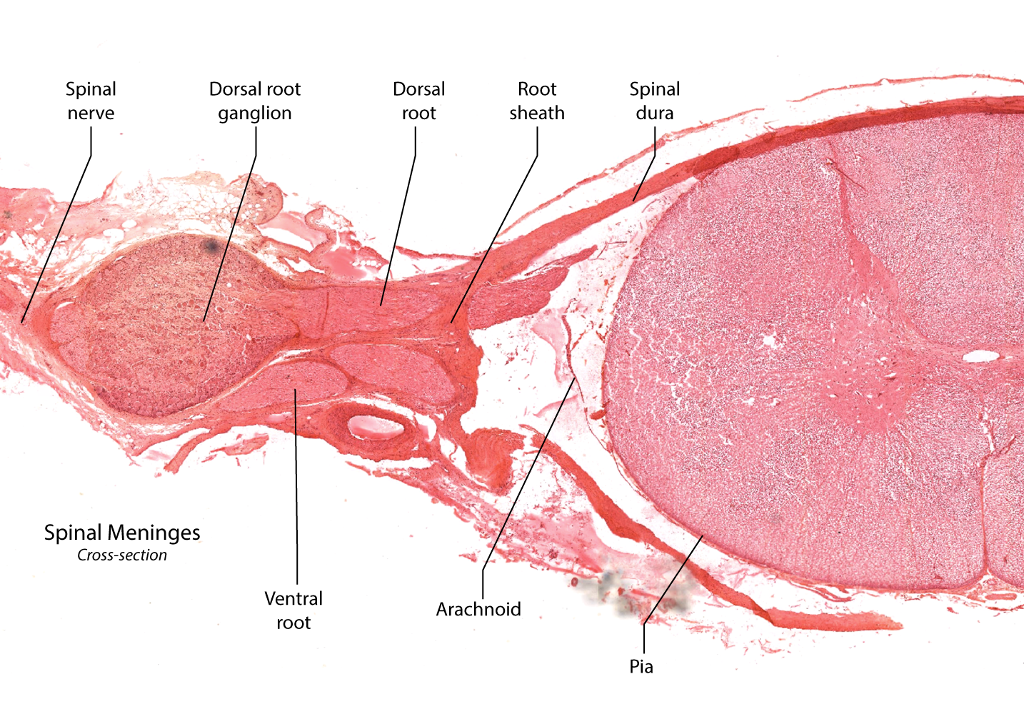
Figure: This is a cross-section of the spinal cord, a nerve root and dorsal root ganglion. Note the dural layer forming a root sheath surrounding the dorsal and ventral roots. |
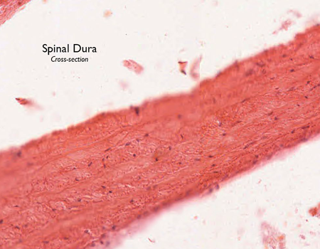 |
Figure: A section of spinal dura. Bundles of collagen fibers are with scattered fibrocyte nuclei. |
Arachnoid
The arachnoid is divided into two layers: a membranous arachnoid that is closely adherent to the inner dural layer and a trabecular arachnoid that is extends inward from the membranous layer to reach the pial layer. The trabeculae are lined with a thin layer of squampus cells. The cerebraospinal fluid (CSF) is contained between the membranous arachnoid and the pial surface. The extracerebral arteries course in the arachnoid before entering brain tissue.
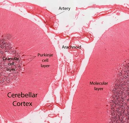 |
Figure: This is a section taken through the folia of the cerebellum. The acrachnoid is seen lying between two of the foloia. An extracerebral artery is seen in the arachnoid. |
 |
Figure: This is a section taken through the cerebral cortex demonstrating two cortical gyri separated by a deep sulcus. Although the membranous arachnoid does not penetrate the sulcus, the trabecular arachnoid does. In this manner the trabeculared arachnoid escorts the extracerebral vessels deep in to the sulcus before these vessels enter the cerebral cortical mantle. |
Pia mater
The pial layer is a thin sheet of collagenous fibers whose surface is lined with a layer of squamous cells. The pial layer is tightly adherent to the surface of the brain or spinal cord.
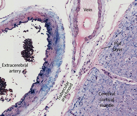 |
Figure: This is an enlargement of the surface of a cerebral gyrus illustration the pial layer on the surface of the cortical mantle and the delicate trabeculated arachnoid escorting the bloods in to the depths of the sulcus. The subarachnoid space surrounds the arachnoid trabeculae and contains the CSF. |
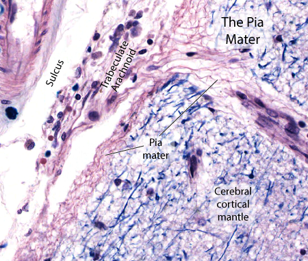 |
Figure: An enlargement of the pial border of the cortical mantle. The pia is seen as a delicate layer of thin collagen fibers lying on the surface of the cerebral mantle. Note how the plial lining follows the blood vessel as it penetrates the cortical mantle. A thin layer of squamous cells form a lining on the arachnoid trabecular as well as on the plial surface. |