Peripheral Nervous System Histology
The peripheral nervous system consists of all structures located distal to the point of fusion between the spinal dura and the epineurium. It is at this point, in the intervertebral canal, that the nerve roots also join together to form the spinal nerves. The peripheral nervous system consists of both nerves and associated ganglia. This section will examine the microstructure of the peripheral nervous system.
Peripheral Nerve
Peripheral nerve arise in the intervertebral canal at the junction of the spinal dura and the epineurim. Upon exiting the intervertebral canal these nerves course throughout the body. The following images represent a view of the cervical and upper thoracic peripheral nerves forming the brachial plexus from which the terminal branches will innervated the upper extremity. Regardless of location location in the plexus or terminal branches, the microstructure of the nerves are similar.
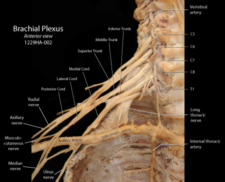 |
Figure: This is an anterior view of the brachial plexus following the removal of all anterior musculature as well as the anterior wall of the thorax. The components of the pleaxus as well as its inital roots and terminal branches all share similar microstructure. |
The figure below is the result of taking a section at right angle to the long axis of the nerve (cross-section). It illustrates multiple nerve fascicles surrounded by a common sheath composed of epineurium.
Epineurium
The epineurial layer is continuous at its origin with the dura mater of the central nervous system. Epineurium is composed of dense connective tissue containing collagen fibers and scattered fibroceytes. The epineurium is divided into an outer layer that surrounds the whole nerve, termed the epidascicular epineurium and an inner layer that surrounds each fascicle with concentric rings, the interfascicular epineurium. Typically the epifascicular epineuroium contains adipose tissue and blends into the surrounding fascial structures that are termed the adventitia of the nerve. The adipose tissue gives each nerve fascicle a degree of movement within the nerve itself. The epineurium is well vasculated, which is termed the vasa nervorum. These blood vessels play an important role in the maintenance of the nerve. Loss of the vasa nervorum, such as occurs in long-standing diabetes, can result in peripheral neuropathy. In addition to fibrocytes, the epineurium contains a varying number of macrophages and mast cells. Finally, the epineurium contains a fine lymphatic plexus and associated lymph vessels, not seen in the nerve fascicles. These vessels drain the epineurium into surrounding lymph nodes.
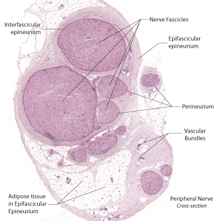 |
Figure: This is a cross-section of a peripheral nerve such as seen in the brachial plexus or its terminal branches. Epifascicular epineurium surrounds the entire structure. Interfascicular epineurium forms a band of concentric rings around each nerve fascicle. |
Perineurium
Surrounding each fascicle in the nerves is a densely-staining but thin sheath composed of multiple wraps of flattened squamous-like cells. Some larger fascicles can have up to ten layers of squamous-like cells in the perineurium. Although the flattened cells appear similar to squamous epithelial cells, they are derived from fibrocytes. The borders of these cells interdigitate with each other and feature zonula occludens and gap junctions supporting the concept that the perineurium forms a permeability barrier around the nerve fascicle.
The perineurim represents the blood-nerve barrier. Thus the perineurium protects the endoneurial space from the epineurial fluids. Since the perineurium creates a tight barrier around the endoneurial space, a rise in pressure in the endoneurial space, termed edema, will result in rising pressures inside the perineurim. The increase pressure can interfere with action potential conduction on individual fibers in the endoneurial space contributing to the signs and symptoms of peripheral neuropathy.
In the histological section below, the perineurium forms a thin, darkly stained band separating the interfascicular epineurium from the nerve fascicle.
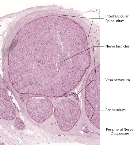 |
Figure: This is a cross-section of several fascicles in a peripheral nerve. The interfsacicular epineurium is seen as a wrapper of dense connective tissue surrounding the fascicles while the perineurium is present as a densly-staining thin sheath of squamous-like cells surrounding each fascilcle. The epifascicular epineurium forms a loose wrapping surrounding all of the nerve fascicules and contains musch adipose tissue. |
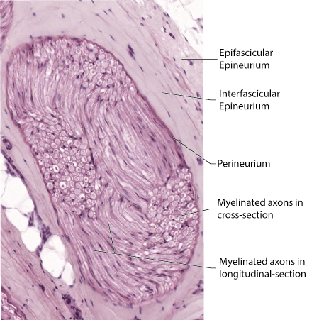 |
Figure: This is an enlargement of a nerve fascicule illustrating the epifascicular and interfsacicular components of the epineurium and the underlying perineurial sheath. The axons present in the nerve fascicule are wavey and have been sectioned in both longitudinal and cross sectional views. |
Endoneurium
Delicate slips of connective tissue extend inward from the perineurium to surround the individual fibers present in the nerve fascicle. This support tissue is termed endoneurium.
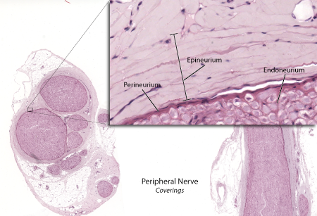 |
Figure: This is an enlargement of the border of a nerve fascicule. The perineurium is seen as a thin, densely staining band separating the interfascicular epineuroium from the nerve fascicule. Within the fascicle delicate strands of endoneurium can be seen separating individual nerve fibers (axon plus Schwann cell sheath). |
The image below is a drawing of a histological section of a peripheral nerve. The interfascicular epineurium is seen as loose bands of connective tissue surrounding the whole nerve while the perineurium is seen as tighter bands of squamous-like calls surrounding a given fascicle. The connective tissue of the perineurium is arranged as a series of tightly organized, flattened lamellae surrounding the fascicle. Perineural septae, termed intrafascicular perineurium, can partition a nerve fascicle as seen in this diagram. Endoneurium is composed of delicate strands of connective tissue penetrating into the individual nerve fascicles and surrounding the nerve fibers.
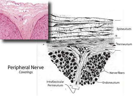 |
Figure: (Figure modified from P. K. Thomas, C.-H. Berthold, and J. L. Ochoa. Microscopic anatomy of the peripheral nervous system: nerve trunks and spinal roots. In: Peripheral Neuropathy, edited by P. J. Dyck and P. K. Thomas, Philadelphia:W.B. Saunders Comapany, 1993, p. 28-91. Original line drawing by Key, A., and Retzius, G., Studein in der Anatomie des Nervernsystems und Bindegewebes, Stockholem, Samson & Wallin, 1876.) |