The Ganglia of the Peripheral Nervous System
| Craniospinal (Dorsal Root) Ganglia | Autonomic Nervous System Ganglia |
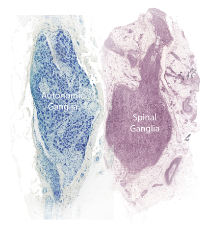
The cell bodies of the motoneurons that innervate skeletal muscle are located in the ventral horn of the spinal cord, however, all other peripheral nerve fibers (axons) have their cell bodies located outside of the central nervous system in clusters termed ganglia. This includes the sensory fibers that are conducted over cranial nerves and spinal dorsal roots as well as the fibers that contribute to he autonomic nervous system. The sensory neurons, regardless of whether that arise in somatic or visceral tissue will have their cell bodies located in the dorsal root ganglia or in ganglia attached to a cranial nerve as it exits the skull; these are termed craniospinal ganglia. The ganglia of the autonomic nervous system are much more varied in location and organization. Some ganglionic neurons are located in the sympathetic trunk that lies along the lateral aspect of the vertebral column while others lie in clusters surrounding the major arteries of the abdominopelvic cavity. Still others form a network that can be found in the walls of visceral organs in the thoracoabdominopelvic cavity.
Craniospinal (Cranial nerve & Dorsal Root) Ganglia
The sensory ganglia of the spinal neurons are located along the dorsal roots positioned inside the root sleeve and inside the intervertebral foramen. These ganglia contain the cell bodies of the sensory neurons that carry impulses from the peripheral tissue to the dorsal horn of the spinal cord and the dorsal column nuclei located at the cervicomedullary junction.
The sensory ganglia contain large pseudounipolar neurons. Each neuron contains a prominent, round nucleus containing a darkly-staining nucleolus. Sensory neurons are arranged in clusters separated by fascicles of nerve fibers and each neuron is surrounded by a capsule of satellite cells. These latter cells are derived from neuroectoderm and are related to Schwann cells. There are no synaptic connections on the sensory neurons contained within sensory ganglia.
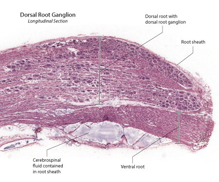 |
Figure: This is a section taken through a the dorsal and ventral roots at the level of the dorsal root ganglion. The entire structure is surrounded by a nerve root sheath. The dorsal root is seen above whilst the ventral root is seen below. The root sheath is composed of dura and arachnoid. A small amount of CSF is seen under the ventral root. The dorsal root ganglion neurons are seen as rows of darkly-staining blobs, separated by sheets of myelinated and unmyelinated axons. |
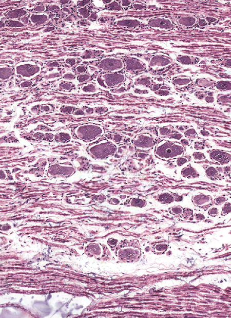 |
Figure: The dorsal root ganglion is composed of numerous ganglion neurons separated by fascicles of nerve fibers. Each ganglionic neuron is pseudounipolar in structure and surrounded by a capsule of satellite cells. |
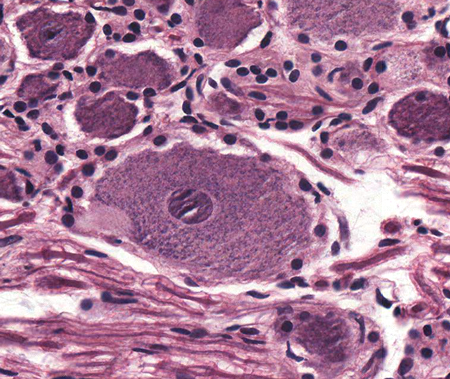 |
Figure: This is a magnified view of a pseudounipolar neuron in the dorsal root ganglion. The neuron has a granular cytoplasm and a large nucleus containing a darkly staining nucleolus. The sensory neuron is surrounded by satellite cells. These later cells are neuroectodermal in origin ans are related to Schwann cells. |
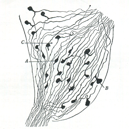 |
Figure: A camera lucida drawing of a dorsal root ganglion stained to demonstrate neuron cell bodies and their axons. Note the psudounipolar nature of most of these sensory neurons. (Figure taken from: Ranson,S.W. and Clark,S.L., The Anatomy of the Nervous System, W.B. Saunders Co., Philadelphia, 1961.) |
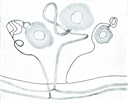 |
Figure: Drawings of the pseudo-unipolar sensory neurons in the dorsal root ganglion. The axons is seen arising from the cell body, coiling several times in the vicinity of the cell body, then joining a fascicle of nerve fibers where it bifurcates. |
| Dorsal Root Ganglia | Autonomic Nervous System Ganglia | Return to the top of the page |
Autonomic Nervous System Ganglia
The ganglia of the autonomic nervous system have extremely varied location and structure. A prominent trunk of sympathetic ganglia is seen in the coursing along the lateral aspect of the vertebral column from the cranial base to the tip of the coccyx at the distal tip of the sacrum. Additional sympathetic ganglia are located in cluster lying along the midline, anterior to the vertebral column and surrounding the root branch of the major abdominopelvic arteries.
Autonomic ganglion typically contain globular-shaped, multipolar neurons, each neuron having several dendrites. The dendrites and the cell bodies of the ganglionic neurons receive synaptic connections from preganglionic fibers. Satellite cells are present in the ganglia but are not organized into the distinct capsules seen in the sensory ganglia.
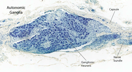 |
Figure: This is a section taken through an ganglion of the autonomic nervous system such as the celiac or the mesenteric ganglia of the abdomen. It features a connective tissue capsule that blends with the surrounding fascia and contains clusters of neurons separated by lightly myelinated and unmyelinated fibers. |
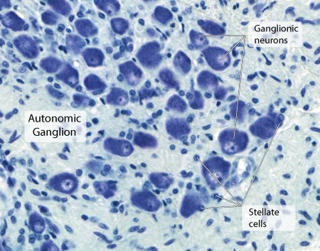 |
Figure: This is a magnified view of the ganglionic neurons. Such cells have large globular-shaped bodies with an eccentric nucleus that has a prominent darkly-staining nucleolus. Satellite cells are present around the neurons but do not form the distinct rings that are seen surrounding the neurons of the sensory ganglia. |
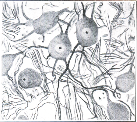 |
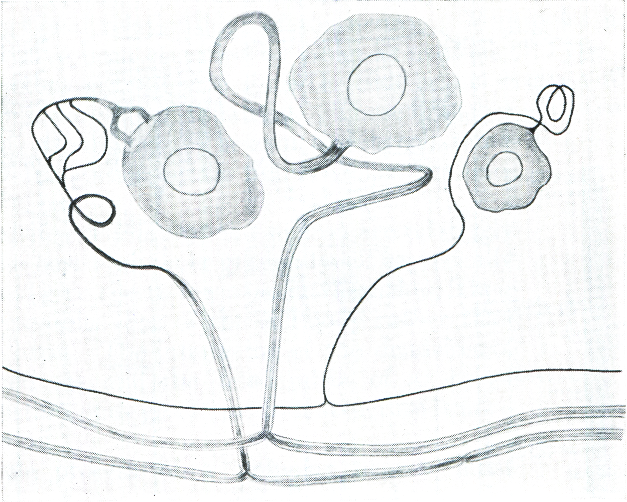
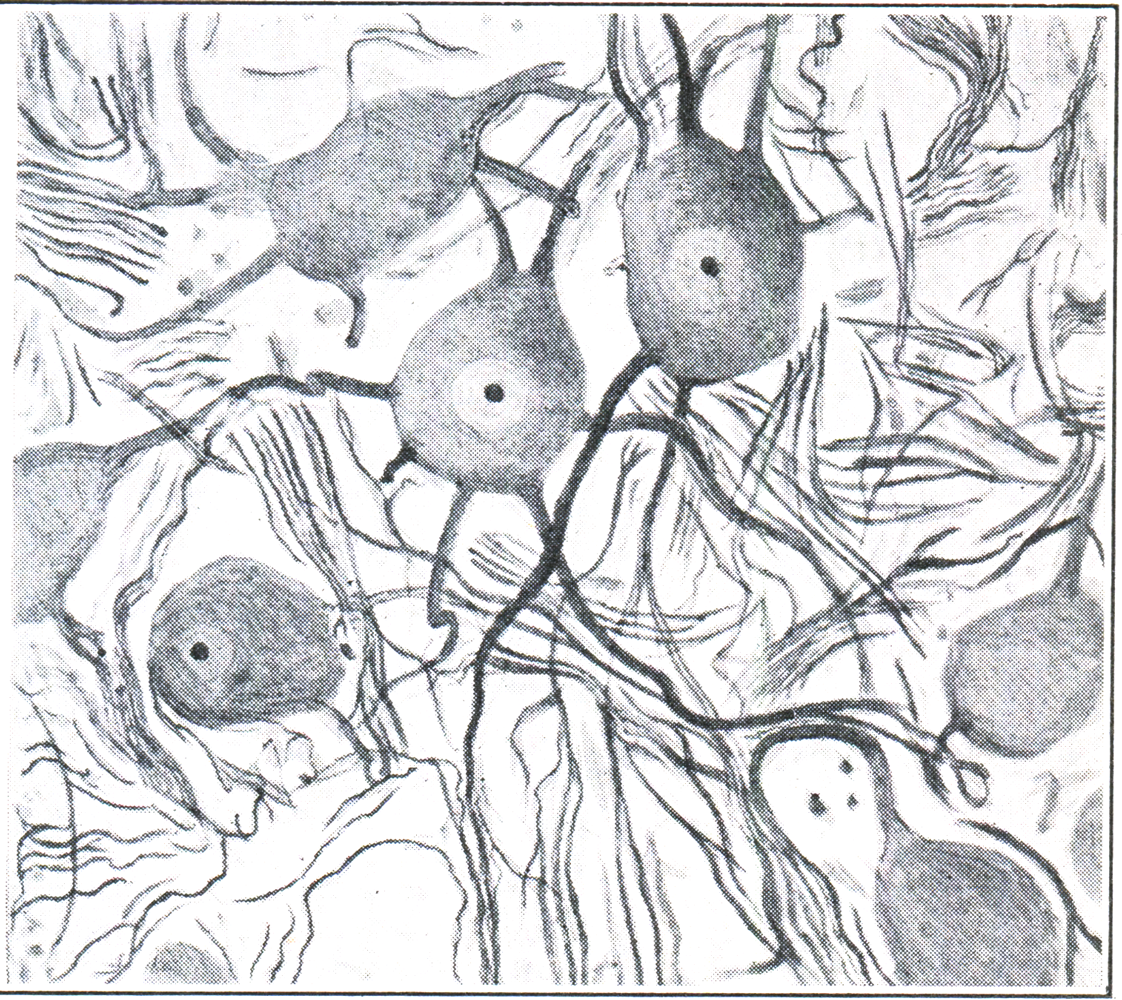
Figure: A drawing of autonomic ganglionic neurons. Although these cells have globular-shaped bodies, they have multiple dendrites making them functionally multipolar cells. The cell bodies and the dendrites receive numerous synaptic endings from the preganglionic fibers that enter the ganglion. |
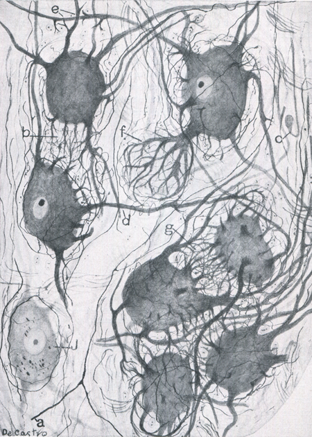 |
Figure: This is a drawing of sympathetic ganglionic neurons taken from a 38-year-old male. (Figure taken from:Larsell, O., Anatomy of the Nervous System, Appleton-Century-Crofts, Inc. New York, 1951) |
| Dorsal Root Ganglia | Autonomic Nervous System Ganglia | Return to the top of the page |