Spinal Cord Microarchitecture
| Dorsal Horn | Lateral Horn | Ventral Horn | White Matter |
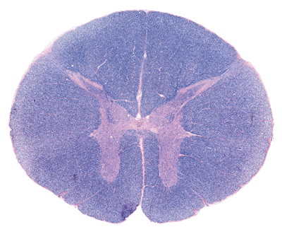
The spinal cord consists of a a central portion of gray matter dominated by neurons and fibers and a surrounding rind of longitudinally-oriented myelinated fibers termed white matter. In the above image of the thoracic spinal cord, the gray matter has and H-shaped distribution been stained a redish color; whilst the surrounding white matter has taken on a blue stain.
The gray matter of the spinal cord and brain is rich in neuronal cell bodies, supporting cells termed neuroglia (protoplasmic astrocytes), and neuronal processes such as axons and dendrites. In a fresh cut specimen this region has a gray appearence. The white matter typically lacks neuronal cell bodies but is rich in axons, both myelinated and unmyelinated, and their support cells the oligodendrocytes and the fibrous astrocytes.
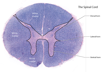 |
Figure: A cross-section of the thoracic spinal cord. The black outline separates the gray matter from the white matter. In this staining technique, the white matter appears blue and the gray matter has a redish color. In the thoracic and upper lumbar regions the gray matter has three horns: dorsal , lateral and ventral. In the remaining portions of the spinal cord only two horns are present: dorsal and ventral. |
Dorsal Horn
The dorsal horn receives sensory information from the primary afferent fibers of the peripheral nerves. Its neurons, termed secondary sensory neurons form axons that project of other regions of the spinal cord and to the brainstem and thalamus. Along with the large sensory neurons, there are numerous smaller interneurons and supporting neuroglial cells.
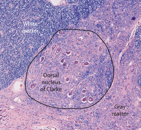 |
Figure: An enlarged view of the base of the dorsal horn to illustrate the clustering of neurons together to form a "nucleus". |
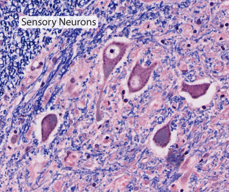 |
Figure: |
Lateral Horn
A small triangular tuft of gray matter extends laterally from the junction of the dorsal and ventral horn in the thoracic and upper lumbar regions of the spinal cord. Contained within the lateral horn are the preganglionic neurons for the sympathetic nervous system.
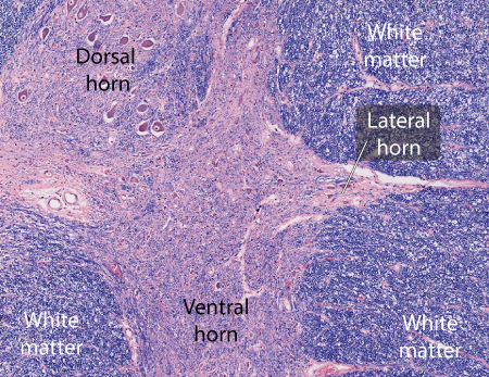 |
Figure: This is an axialplane section taken through the junction of the dorsal and ventral horn in thoracic spinal cord. The lateral horn is a triangular tuft of gray matter extending laterally from this junction. |
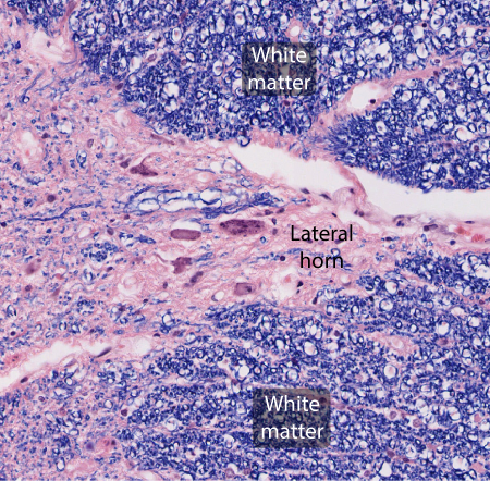 |
Figure: An enlarged view of the lateral horn illustrating several large oval preganglionic neurons. |
Ventral Horn
The ventral horn is a thick mass of gray matter containing the motoneurons that innervated the skeletal muscles of the body. The largest of the motoneurons, the alpha-motoneurons, are the largest cells of the spinal cord. These cells are surrounded by smaller gamma-motoneurons, interneurons and numerous neuroglial cells.
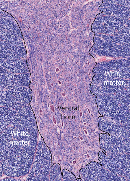 |
Figure: This is an axial plane section taken through the ventral horn in the thoracic region. |
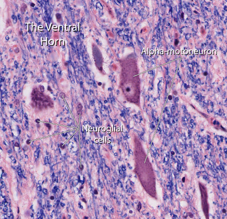 |
Figure: This is an enlarge view of the ventral horn demonstrating the alpha-motoneuron cell bodies. |
White Matter
The white matter forms a thick rind surrounding the spinal cord. It is composed of many ascending and descending axons and their supporting sheaths. Rays of transverse fibers are also seen passing in and out of the dorsal and ventral horns. Two types of neuroglia form the principal support cells of the white matter: oligodendrocytes and protoplasmic astrocytes. The oligodendrocytes provide the myelin sheaths for axons while the astrocytes serve as part of the blood-brain barrier.
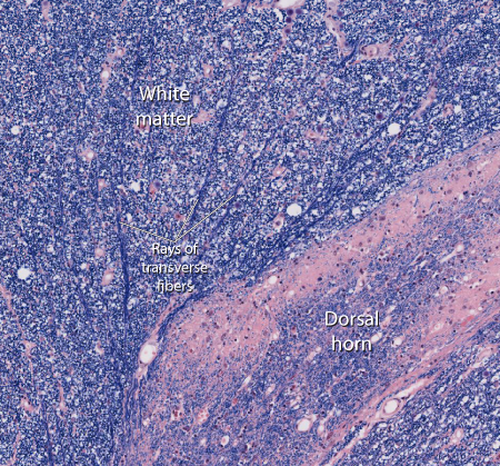 |
Figure: This is an axial plane view of the border between the dorsal horn and the surrounding white matter. Several rays of transverse fibers are seen approaching the medial border of the dorsal horn. |
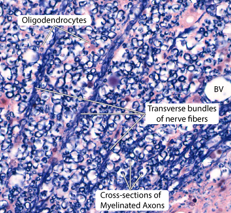 |
Figure: An enlarged view of the white matter demonstrating cross-section and transverse bundles of axons along with oligodendroglial cells. |