Ventral View of the Brain Stem
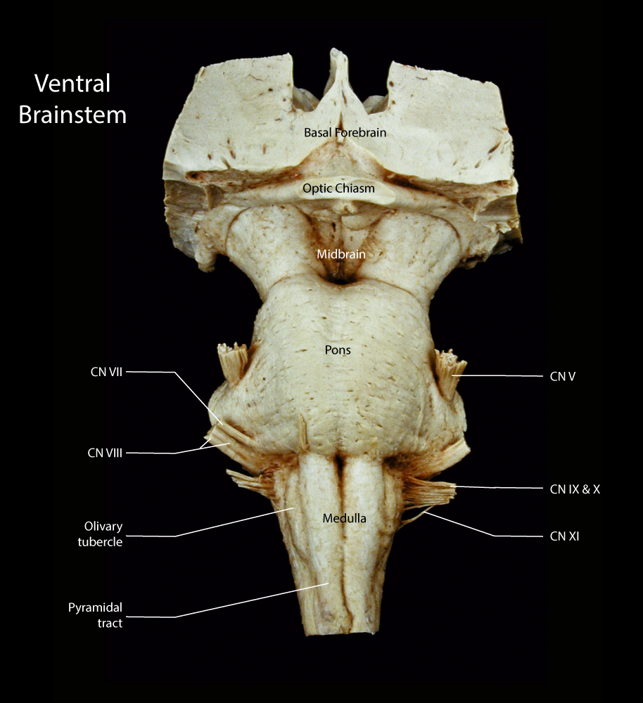
The ventral view of the brainstem features the cerebral peduncles in the midbrain, the cerebellar peduncle in the pons and the pyramidal tract in the medulla.

A ventral view of the brainstem
Enlarge this image in a new window
The cerebral peduncles contain the axons of cortical neurons descending into the brainstem and spinal cord. These fiber bundles disappears into the pons which contains massive bundles of axons arising in pontine nuclei and extending into the cerebellum. From the distal pons the corticospinal axons form the pyramidal tracts. Lateral to the pyramidal tracts is an eminence, the olivary tubercle, created by the inferior olivary nucleus. This later structure represents a relay nucleus with projections into the cerebellum.
Cranial nerve twelve emerges from the brainstem in the groove between the pyramidal tract and the olivary tubercle. Dorsal to the tubercle, the roots of cranial nerves 9-11 emerge from the brainstem. At the pontomedullary junction cranial nerves seven and eight emerge laterally while six emerges ventrally. From the center of the pons the large root of cranial nerve five is seen. Finally, cranial nerve three emerges from the medial aspect of the cerebral peduncles in the midbrain.