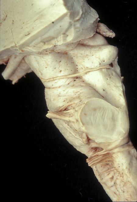
Lateral View of the Brainstem
The brainstem represents an elongation of the spinal cord. The changes in shape seen in the brainstem are due to the addition of nuclei and circuits controlling the cranial nerves and the pathways accessing the cerebellum as well as the expansion of the gray matter core to house the regions controlling cardiorespiratory and other homeostatic functions in the body.
Medulla
In the lateral view the medulla presents the olivary tubercle created by the enlarged inferior olivary nucleus, a cerebellar relay nucleus. Cranial nerves VII, VIII, IX, X, and XI are seen leaving the brainstem in a sulcus bordered by the olivary tubercle ventrally and the inferior cerebellar peduncle dorsally.
Pons
The massive bulge of the pons, created by the pontine nucleus, extends dorsally to form the middle cerebellar peduncle. Toward the rostral border of the middle cerebellar peduncle, cranial nerve V leaves the brainstem.
Midbrain
The lateral view of the midbrain presents to distinctive features. Ventrally, the cerebral peduncle, the pathway for the corticospinal and corticonuclear fibers is seen emerging from the forebrain to disappear into the pons. Dorsally the superior and inferior colliculi are seen rising upward between the thalamus and the cerebellum. The superior colliculus is a relay for visual reflexes and the inferior colliculus is a relay for the ascending fibers of the auditory system.
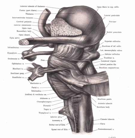 |
Figure: This is a lateral view of the brainstem and thalamus. The globular-shaped thalamus is located superior to the brainstem. |