Dorsal View of the Brain Stem
The dorsal surface of the brainstem can only be visualized by removing the cerebellum as has been done in Figure 01. This procedure exposes the pontomedullary junction as well as the floor of the fourth ventricle. This ventricle is rhomboid in shape, at its widest points it is bounded by the middle cerebellar peduncles. Superior to the ventricle are the inferior and superior colliculi.
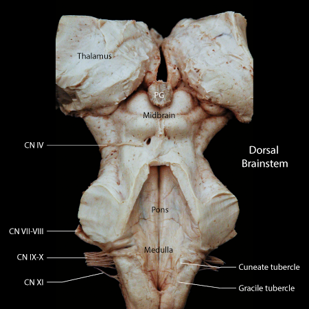
Figure 01: A dorsal view of the brainstem following the removal of the cerebellum
Medulla
The medulla extends from the cervical spinal cord to the inferior margin of the middle cerebellar peduncle. The transition from cervical spinal cord to medulla has few external features, conversely at the rostral end of the medulla, the junction with the pons is abrupt. The dorsal aspect of the medulla in dominated by the wedge shaped opening of the fourth ventricle. The ridges on either side of the ventricle contain two tubercles, the gracile tubercle and the cuneate tubercle. Each tubercle contains the respective nucleus such that the cuneate tubercle contains the nucleus cuneatus as does the gracile tubercle contain the nucleus gracilis. Each nucleus receives synaptic endings from its respective fiber tract.
At the rostral extent of the medulla a narrow raise band of tissue wraps around the dorsal ridge just inferior to the middle cerebellar peduncle. This band is created by the dorsal cochlear nucleus, a component of the auditory pathways. Four cranial nerves are seen exiting the medulla, from caudal to rostral, the hypoglossal nerve (not seen in the dorsal view) is the caudal most nerve to exit the medulla, its root is close the the midline on the ventral side of the brainstem, however, the hypoglossal nucleus forms a bulge located close to the midline in the caudal portion of the floor of the fourth ventricle, thus the hypoglossal tubercle can be seen in the dorsal view. Laterally, contributions to the cranial nerve eleven, ten and nine exit the medullary portion of the brainstem.
Pons
The structure of the pons is dominated by the connections to the cerebellum, in fact, the pons represents a relay for axonal projections from vast regions of the cerebrum to the cortex of the cerebellum, a pathway termed the Cortico-Ponto-Cerebellar tract. The collection of neurons relaying this tract to the cerebellum, the pontine nucleus, is very large resulting in the bulbous expansion of this region of the brainstem. The nuclei for several cranial nerves are contained within the pons. The vestibular nuclei and the cochlear nuclei are found clustered around the entrance of the eighth cranial nerve. Parallel to CN 8 lies the facial nerve (CN. VII). The nucleus of cranial nerve seven is situated deep in the caudal pons. its axons pass dorsally to curve over the nucleus of cranial nerve six and then laterally to leave the brainstem. Ventrally, cranial nerve six leaves the brainstem in close juxtaposition to the corticospinal tract, however, the nucleus of cranial nerve six is located dorsally in the brainstem and forms an eminence on the floor of the fourth ventricle.
Midbrain
The dorsal view of the midbrain is dominated by the superior and inferior colliculi. A large bundle of axons, termed the brachium (arm) of the inferior colliculus, leaves this structure and passes superiorly toward the body of the thalamus. This fiber bundle contains auditory information in route through the thalamus to the auditory portion of the cerebral cortex. Just below the inferior colliculus the fourth cranial nerve is seen emerging from the brainstem. This nerve is the only cranial nerve to arise from the dorsal surface of the brainstem. The other cranial nerve in the midbrain, the oculomotor nerve, is located on the ventral border of the cerebral aqueduct and its axons leave the brainstem ventrally.