Spinal Cord Organization
The internal structure of the spinal cord is divided into two major compartments, a core of gray matter composed on neurons, neuroglia and their processes and a surrounding rind of white matter composed of myelinated axons and their supporting neuroglia. The white matter rind diminishes in thickness along the length of the spinal cord from cervical to coccygeal segments. The gray matter changes in thickness according to the function of this region. Thus the gray matter is thickest in the lower cervical region C5-T1 (reflecting the brachial plexus and upper extremity) and in the lower lumbar and upper sacral region L4-S3 (reflecting the lumbosacral plexus and the lower extremity). Conversely the gray matter is thinnest in the upper cervical region (innervating the neck) and in the thoracolumbar region (innervating the torso).
Gray Matter
Dorsal horn - The dorsal horn receives sensory information from the central processes of the primary afferent fibers. It processes this information and elicits two major outcomes. The initiation of spinal reflexes to adjust the position of the associated limbs and the relaying of sensory information upstream to the brainstem and thalamus where it can ultimately reach the level of the cerebral cortex. The dorsal horn is divided into three regions. A small cap of neurons sits on the dorsal most portion of the horn, termed the posterior marginal nucleus. Next, a curved band of neurons forms the substantia gelatinosa. this later nucleus wraps around the dorsal aspect of the largest structure in the dorsal horn, the nucleus proprius. The three nuclei are involved in the processing of nociceptive (pain) information from primary afferent fibers.
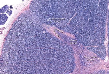
Figure: A section of the thoracic dorsal horn illustrating the nucleus posteriomarginalis, nucleus proprius and dorsal nucleus of Clarke
Ventral horn - This column of gray matter extends the full length of the spinal cord. It contains the large motor neurons whose axons form the ventral rootlets and innervate the skeletal muscles of the body. It varies in thickness along the length of the spinal cord being widest in the lower cervic al and lumbosacral regions and narrow in the upper cervical and thoracic and upper lumbar regions. Essentially the two areas of expasion represent the brachial and lumbosacral plexi and reflect the need for more neurons to innervate the muscles in the upper and lower extremity.
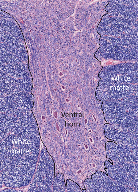
This is a view of the ventral horn taken from a coronal section through the thoracic spinal cord. Th elarge cells distributed in clusters represent alpha-motoneurons. Their axons exit the spinal cord through the ventral rootls and directly innervated skeletal muscle in the trunk.
Within each of the spinal enlargements the ventral horn adds a lateral column of gray matter containing motoneurons and associated interneurons. Within the lower cervical spinal cord (C5-T1) the epansion represents the upper extremity. The arrangement of motoneurons across the entire cervical ventral horn is reflective of a body map. The neurons innervating trunk musculature are located most medially whilst those innervating the musculature controling the digits are located most laterally in the lateral expansion. The representation of the proximal limb muscles is located in between these two extremes. Generally the flexor muscles are represented dorsally and the extensor muscles ventrally. This type of orderly organization is termed a somatotopic map and is typical of the way in which the central nervous system represents both motor and sensory information.

This is a coronal section taken through the cervical enlargement.
Enlarge this image in a new window
The transition from the cervical enlargement into the thoracic spinal cord involves the loss of the lateral expansion of the ventral horn. The remaining ventral horn simply represents the trunk musculature present in the thoax and abdomen. Around L3 the lateral expansion re-occurs and represents the lower extremity in a topographic manner similar to that seen in the upper extremity. The lower extremity expansion extends from L3 to S3 or S4). The representation of muscles controling the bladder and bowel sphincters are located in the medial portion of the lower aspect of this expansion (S3-4).
Lateral horn - From spinal cord segments T1 to L2 or L3 a small lateral epansion exists between the dorsal and ventral horns. This lateral horn contains the representation of the sympathetic preganglionic neurons.
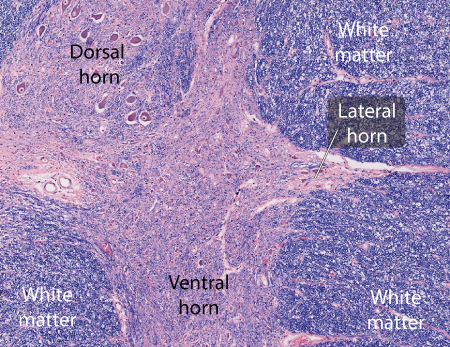
A coronal section of the midthoracic spinal cord demonstrating the location of the lateral horn inbetween the dorsal and ventral horns
Axons from the neurons located in the lateral horn pass downward through the ventral horn and enter the ventral roots with the axons from the alpha-motoneurons. The ventral root then merges with the dorsal root in the intervertebral foramen to form a spinal nerve. Shortly after the spinal nerve exits the intervertebral foramen, these preganglionic axons leave the nerve on a white ramus leading to the sympathetic trunk ganglia on the posterior body wall.
Commissural or Intermediate Region - The two masses of gray matter in the spinal cord are connected across the midline by a narrow band of gray matter termed the commissural or intermediate region. Contained within this commissure is the central canal of the spinal cord. This structure is patient in the new born and contains some cerebrospinal cord, however by adulthood the canal os obliterated in some portions of the spinal cord. Lying along the ventral border of the gray commissure is a white commissure composed of the decussating of spinal cord neurons. Most of these axons originate from dorsal horn neurons and contribute to the anterolateral or spinothalamic tract in the white matter of the spinal cord.
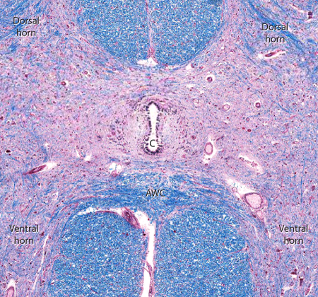
A coronal section through the lombosacral enlargement demonstrating the gray and white commissure of the spinal cord. The central canal (C) is seen in the center of the white matter while the anterior white commissure (AWC) composed of decussationg axons lies ventral to the gray commissure.
White Matter
The white matter, composed of myelinated and unmyelinated axons, forms a rind surround the gray matter of the spinal cord. These fibers are of three types: 1) long ascending fibers that arise in the spinal cord and extending into the brainstem and thalamus, 2) long descending fibers that arise in the cerebral cortex or brainstem and terminate at various levels on the spinal cord, and 3) propriospinal axons that arise at one level of the spinal cord and terminate in another level but never leave the spinal cord.
The rind of white matter is subdivided by region such that four funniculi are recognized:
1) Dorsal funniculus
2) Dorsolateral funniculus
3) Ventrolateral funniculus
4) Ventral funniculus.
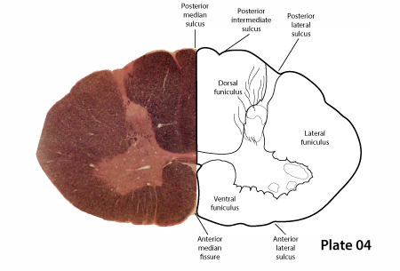 |
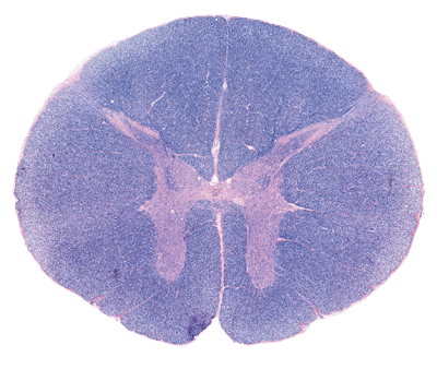
Figure: This is a coronal section of the lower cervical spinal cord indicating the major surface features of the cord. The dorsal rootlets enter the spinal cord through the posterior lateral sulcus, while the ventral rootlets leave the cord through the anterior lateral sulcus. The posterior lateral sulcus is deeper than the anterior lateral sulcus. The anterior median fussure is a true space, extending into the spinal cord as deep as the gray grsy matter. |
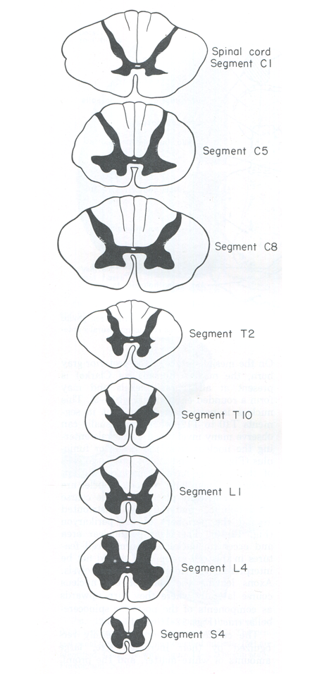 |
Figure: This is a schematic diagram of the 8 spinal cord segments progressing from cervical to sacral levels. It illustrates the change in shage of the spinal cord as well as its gray and white matter along the long axis of the cord. Note that the posterior intermediate septum divides the dorsal funniculus into the fasciculus grasilis and cuneatus in the upper thoracic and all cervical segments. This septum first appears at the T6 level. |