The Gross Anatomy of the Spinal Cord
Coverings of the Spinal Cord
The spinal cord is an elongated column of white and gray matter that lies wrapped in three layers of meninges and suspended inside the bony walls of the vertebral (spinal) canal. The outermost layer is dura and composed of dense connective tissue. Inside the dural layer is a thin velum or membrane of arachnoid and on the surface of the spinal cord is a layer of pial cells. Between the membranous arachnoid and the pia there exist numerous, delicate trabeculae of arachnoid. It is in this subarachnoid space that the cerebrospinal fluid is located. Laterally, the pial layer extends off the margin of the spinal cord forming the denticulate ligament. The lateral margin of this ligament is scalloped and forms 31 points, each point penetrating through the membranous arachnoid to attach to the dura. The denticulate ligaments stabilize the spinal cord within the meningeal layers. Dorsal (sensory) and ventral (motor) rootlets arise from the lateral aspect of the cord, several rootlets unite to form a root and the dorsal and ventral roots pass around the denticulate ligament into the lateral recess in the dura sac finally entering the dural root sleeve. The dorsal root ganglion is located in the root sleeve, distal to this structure the dorsal and ventral roots merge to form a spinal nerve.
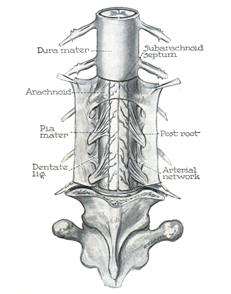
The spinal cord and its surroundings (Figure taken from: P. Thorek. Anatomy in surgery, New York:Springer-Verlag, 1985.)
During development the spinal cord and the spinal canal are similar lengths, however the vertebral column grows faster than the cord, thus a length discrepancy emerges between the two structures. At 20 weeks of gestation, the differential growth rate of the spinal cord and vertebral column has placed the distal end of the cord, termed conus medullaris, at the L4-L5 level of the column. By 40 weeks gestation the conus is located at the L3 level and by 2 month of postnatal life the conus is located at the L1-L2 level of the column, which is the adult position. Although the spinal cord ends at the L1-L2 vertebral level, its roots continue to descend ultimately reaching their appropriate root sleeve in the lower lumbar and sacral vertebral levels. The dural sac and the arachnoid continue downward to eventually close off at the level of the S2 in the sacrum. The configuration of a spinal cord narrowing to a termination at L1 and long lumbar and sacral root extending below gave rise to the term cauda equina - the "horse's tail".
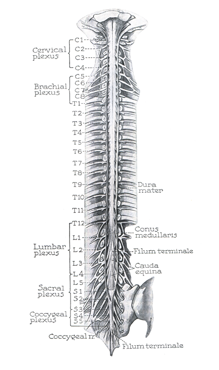
This is an illustration demonstrating the full extent of the spinal cord in the dural sac. (Figure taken from: P. Thorek. Anatomy in surgery, New York:Springer-Verlag, 1985.)
Shape of the Spinal Cord
The shape of the spinal cord reflects its function. Proximally, the cord expands to form the cervical enlargement that extends from approximately C5 to T1. There after the cord shrinks in diameter between t T2 and L2 and re-expands between L3 and S3 forming the lumbosacral enlargement. Finally it narrows to a small pointed tip from which a cord of pial, termed the filum terminale extends to the distal tip of the dural sac. The cervical enlargement houses the circuitry necessary for the upper extremity while that for the lower extremity is contained in the lumbosacral enlargement. The tapering of the distal end of the spinal cord is termed the conus medullaris.
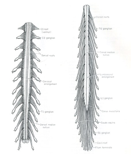
A drawing of the spinal cord illustrating its progressive shape changes along its long axis. (Figure taken from: E. C. Crosby, T. Humphrey, and E. W. Lauer. Correlative Anatomy of the Nervous System, New York:The Macmillan Company, 1962.)
External Features of the Spinal Cord
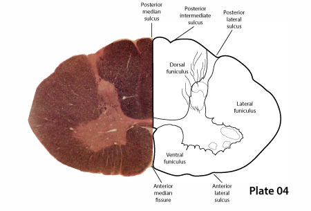
The perimeter of the spinal cod has several external features of note. Dorsally, there is a posterior median septum that separates the two dorsal funiculi on the midline. A posterior lateral sulcus marks the point where the dorsal root entry zone (DREZ) occurs. The dorsal roots carry sensory information into the spinal cord. Ventrally there are two significant feature to the spinal cord. An anterior median fissure cleaves the spinal cord on the midline as far internally as the anterior white commissure. This prominent structure is a true open space and not a septum. Also an anterior lateral sulcus marks the region where the ventral roots emerge from the spinal cord. The ventral roots carry motor information away from the spinal cord. The anterior lateral sulcus is not as prominent as the dorsal lateral sulcus, thus reflects the fact that the dorsal rootlets are far more numerous than the ventral rootlets.
Segmental Organization
The spinal cord typically gives rise to 31 pairs of spinal nerves - 8 cervical, 12 thoracic, 5 lumbar, 5 sacral and 1 coccyx. Based on these nerves, the spinal cord is often thought off as having well defined segments, however, the position of the 31 nerves is determined, not by the genetic segmentation of the spinal cord but by the clustering of nerve rootlets, which is accomplished by the growth of the vertebral arch structures during development. In reality, the spinal cord has a homogeneous expression of genes along its full length and, thus, is not segmented. Segmentation is enforced on the the spinal cord indirectly by the clustering of the nerve rootlets to make specific spinal nerve.
The first spinal nerve is an odd structure, it passes posteriorly over the arch of the C1 vertebra leaving the vertebral canal without going through a intervertebral foramen. It enters the suboccipital triangle and innervates the small muscles that define the triangle; typically this nerve has no sensory component. The second cervical nerve also passes posteriorly to exit the vertebral canal by slipping between the arch of C1 above and the lamina of C2 below, it does not involve an intervertebral foramen in this route. All remaining spinal nerves exit the spinal canal by using an intervertebral foramen.
With C2 spinal nerve passing above the lamina of C2, that leaves C3 to pass between C2 vertebra above and C3 below. This pattern persists until C7. The C7 spinal nerve passes between C6 vertebra above and C7 below and the C8 spinal nerve passes under the C7 vertebra but above the T1 vertebra. This sets up T1 to begin a new pattern with the T1 spinal nerve passing under the T1 vertebra, T2 spinal nerve under the T2 vertebra etc. This latter pattern will persist throughout the remainder of the spinal cord.
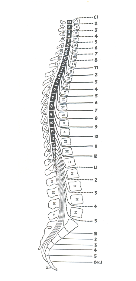
This segmental drawing show the C1 spinal nerve passing over the arch of the C1 vertebra. All remanding cervical nerves pass over their respective vertebra until C8 which passes under the C7 vertebra but over the T1 vertebra. From here on down all spinal nerve exit the spinal canal by passing under their associated vertebral structure. (Figure taken from R. C. Truex and M. B. Carpenter. Human Neuroanatomy, Baltimore:Williams & Wilkins Co., 1969.)
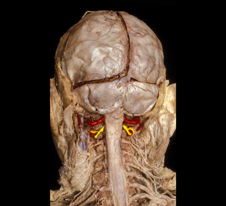 |
Figure:
|
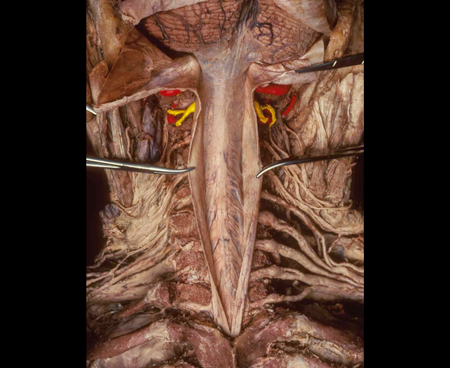 |
Figure: |
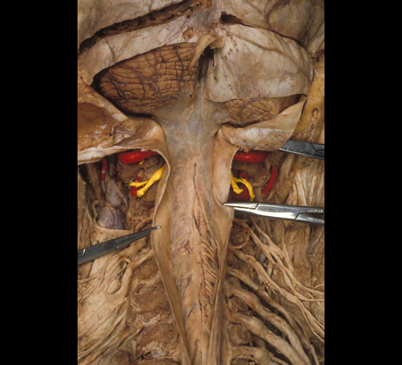 |
Figure: |
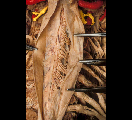 |
Figure: |
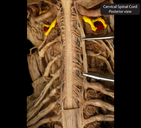 |
Figure: This is a posterior view of the cervical spinal cord in an 80-year-old female following a laminectomy and the removal of the dura and arachnoid. |
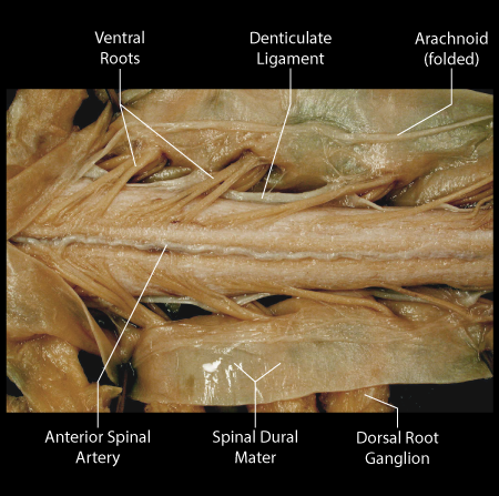 |
Figure: This is a ventral view of the cervical enlargement of the spinal cord. The ventral (motor) rootlets emerge from the ventral lateral sulcus. |
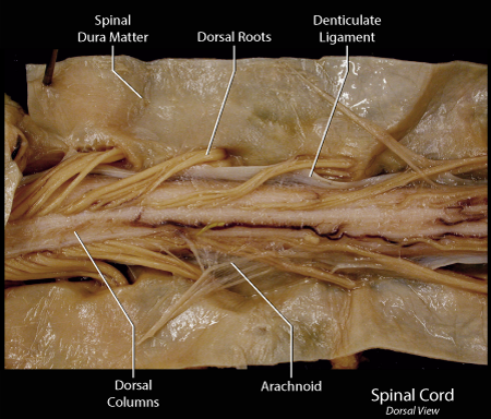 |
Figure: This is a dorsal view of the cervical enlargement of the spinal cord. The dorsal (sensory) rootlets emerge from the dorsal lateral sulcus. |
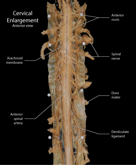 |
Figure: This is an anterior view of the cervical enlargement of the spinal cord. |
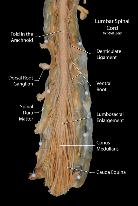 |
Figure: This is an anteriorl view of the lumbosacral enlargement of the spinal cord. |
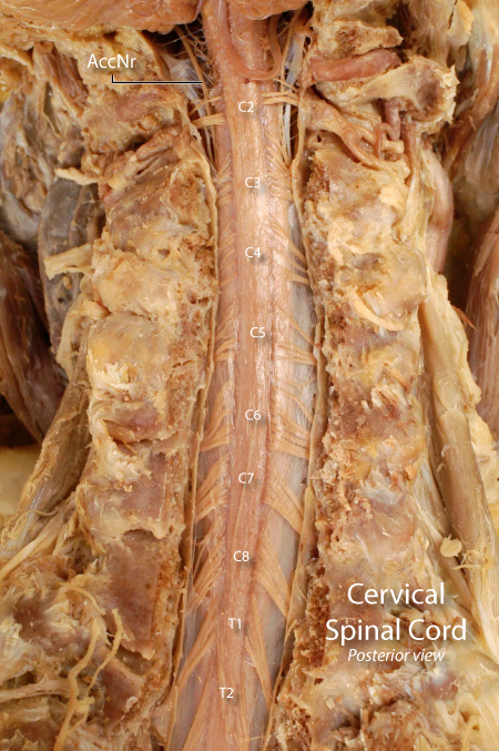 |
Figure: This is a posterior view of the cervical enlargement of the spinal cord. |
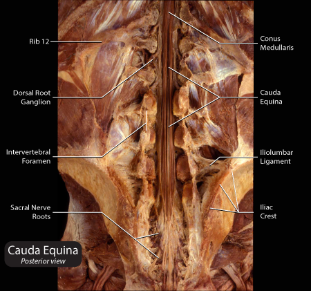 |
Figure: This is a posterior view of the cauda equina. |